Comparative Study
. 2006 Oct 2;203(10):2271-9.
doi: 10.1084/jem.20061308. Epub 2006 Sep 18.

Interleukin (IL)-22 and IL-17 are coexpressed by Th17 cells and cooperatively enhance expression of antimicrobial peptides

Affiliations
Comparative Study

Interleukin (IL)-22 and IL-17 are coexpressed by Th17 cells and cooperatively enhance expression of antimicrobial peptides

Spencer C Liang et al. J Exp Med. .

Abstract

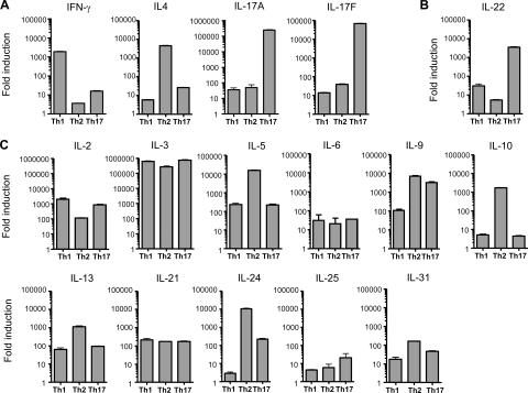
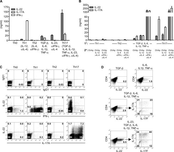
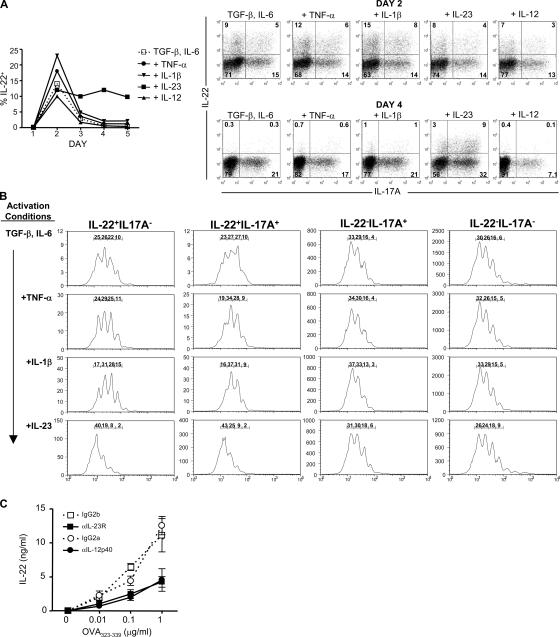
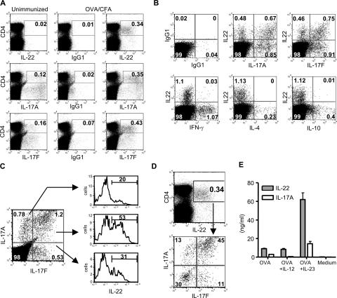
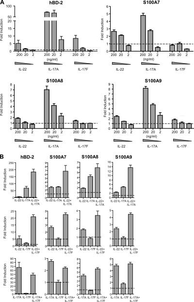
Th17 cells are a distinct lineage of effector CD4(+) T cells characterized by their production of interleukin (IL)-17. We demonstrate that Th17 cells also expressed IL-22, an IL-10 family member, at substantially higher amounts than T helper (Th)1 or Th2 cells. Similar to IL-17A, IL-22 expression was initiated by transforming growth factor beta signaling in the context of IL-6 and other proinflammatory cytokines. The subsequent expansion of IL-22-producing cells was dependent on IL-23. We further demonstrate that IL-22 was coexpressed in vitro and in vivo with both IL-17A and IL-17F. To study a functional relationship among these cytokines, we examined the expression of antimicrobial peptides by primary keratinocytes treated with combinations of IL-22, IL-17A, and IL-17F. IL-22 in conjunction with IL-17A or IL-17F synergistically induced the expression of beta-defensin 2 and S100A9 and additively enhanced the expression of S100A7 and S100A8. Collectively, we have identified IL-22 as a new cytokine expressed by Th17 cells that synergizes with IL-17A or IL-17F to regulate genes associated with skin innate immunity.


Figures

Figure 1.
Figure 1.
IL-22 transcript expression is high in Th17 cells. Purified CD62LhiCD4+ (naive) DO11 T cells were activated with irradiated splenocytes, OVA323–339 (OVAp), and various cytokines and antibodies to differentiate cells to Th1 (IL-12, anti–IL-4), Th2 (IL-4, anti–IFN-γ), and Th17 (TGF-β, IL-6, IL-1β, TNF-α, IL-23, anti–IFN-γ, anti–IL-4) lineages. On day 7 of culture, CD4+ T cells were repurified and rested overnight. Cells were then restimulated with PMA and ionomycin under differentiation conditions for 6 h. RNA was prepared and quantitative PCR was performed in A to ensure that cells were properly differentiated. (B and C) ILs known to be expressed in T cells were evaluated by quantitative PCR. All transcript amounts were normalized to HPRT, and fold induction was calculated relative to unactivated, naive DO11 T cells. Data shown are representative of two independent experiments. Error bars are SD.
Figure 2.
Figure 2.
Th17 cells express IL-22 protein. (A) Naive DO11 T cells were activated with irradiated splenocytes, OVAp, and various cytokines and antibodies. IL-22, IL-17A, and IFN-γ concentrations were measured on day 5. (B) Naive DO11 cells were differentiated under Th1, Th2, or Th17 conditions, or with TGF-β, IL-6, IL-1β, and TNF-α for 7 d. After resting overnight, cells were then restimulated with OVAp, IL-2, irradiated splenocytes, and combinations of cytokines and antibodies. IL-22 and IL-17A concentrations were measured on day 5. (C) Intracellular cytokine staining was performed on cells from A on day 5. Plots are gated on KJ126+CD4+ cells. (D) Naive DO11 T cells were activated as in A with the indicated cytokines. Intracellular cytokine staining was performed on day 5. Data shown are representative of three independent experiments. Error bars are SD.
Figure 3.
Figure 3.
IL-23 expands IL-22–producing cells. CFSE-labeled naive DO11 T cells were activated with OVAp, irradiated splenocytes, TGF-β, and IL-6. TNF-α, IL-1β, IL-23, or IL-12 was added to some cultures. Intracellular cytokine staining for IL-22 and IL-17A was performed on days 1–5. (A) Percentages of IL-22+ cells on day 1–5 and representative plots on days 2 and 4 are shown. (B) CFSE profiles on day 4 of cells separated into four populations: IL-22+IL-17A, IL-22+IL-17A+, IL-22IL-17A+, and IL-22IL-17A. Percentage of cells in each peak is shown. (C) Naive DO11 T cells were cultured with LPS-activated DCs, OVAp, and neutralizing antibodies to IL-23R, IL-12p40, or respective isotype controls. IL-22 concentrations were determined on day 5. Data are representative of at least two experiments.
Figure 4.
Figure 4.
IL-22 is coexpressed with IL-17A and IL-17F in vivo. C57BL/6 mice were immunized with 100 μg OVA emulsified in CFA. 7 d after immunization, popliteal LN cells were harvested and the intracellular cytokine staining protocol was immediately performed. (A) LN cells were stained for CD4 and IL-22, IL-17A, and IL-17F. (B) IL-22 expression was analyzed in relation to IL-17A, IL-17F, IFN-γ, IL-4, and IL-10 in CD4+ T cells. (C) Expression of IL-22 in IL-17A+ and/or IL-17F+ populations was analyzed. (D) Expression of IL-17A and IL-17F in IL-22+ cells was determined. (E) LN cells were restimulated with 200 μg/ml OVA. IL-12 or IL-23 was added as indicated. IL-22 and IL-17A concentrations were examined on day 4. Data shown are representative of three independent experiments. Error bars are SD.
Figure 5.
Figure 5.
IL-22 in combination with IL-17A or IL-17F cooperatively enhances the expression of antimicrobial peptides. Primary human keratinocytes were stimulated with IL-22, IL-17A, or IL-17F individually (A) or with pairwise combinations (B) of 200 ng/ml IL-22, 20 ng/ml IL-17A, and 20 ng/ml IL-17F for 44 h. Relative amounts of hBD-2, S100A7, S100A8, and S100A9 transcript were determined by quantitative PCR and normalized to GAPDH. Fold induction was calculated relative to untreated keratinocytes (dashed line). Data are representative of at least two experiments performed on each of three separate donors. Error bars are SD.

Similar articles

Cited by

References

    1. Kolls, J.K., and A. Linden. 2004. Interleukin-17 family members and inflammation. Immunity. 21:467–476. - PubMed
    1. Aggarwal, S., N. Ghilardi, M.H. Xie, F.J. de Sauvage, and A.L. Gurney. 2003. Interleukin-23 promotes a distinct CD4 T cell activation state characterized by the production of interleukin-17. J. Biol. Chem. 278:1910–1914. - PubMed
    1. Langrish, C.L., Y. Chen, W.M. Blumenschein, J. Mattson, B. Basham, J.D. Sedgwick, T. McClanahan, R.A. Kastelein, and D.J. Cua. 2005. IL-23 drives a pathogenic T cell population that induces autoimmune inflammation. J. Exp. Med. 201:233–240. - PMC - PubMed
    1. Harrington, L.E., R.D. Hatton, P.R. Mangan, H. Turner, T.L. Murphy, K.M. Murphy, and C.T. Weaver. 2005. Interleukin 17-producing CD4+ effector T cells develop via a lineage distinct from the T helper type 1 and 2 lineages. Nat. Immunol. 6:1123–1132. - PubMed
    1. Park, H., Z. Li, X.O. Yang, S.H. Chang, R. Nurieva, Y.H. Wang, Y. Wang, L. Hood, Z. Zhu, Q. Tian, and C. Dong. 2005. A distinct lineage of CD4 T cells regulates tissue inflammation by producing interleukin 17. Nat. Immunol. 6:1133–1141. - PMC - PubMed

Publication types

MeSH terms