. 2006 Oct 1;177(7):4376-83.
doi: 10.4049/jimmunol.177.7.4376.

Blockade of CTLA-4 on CD4+CD25+ regulatory T cells abrogates their function in vivo

Affiliations

Blockade of CTLA-4 on CD4+CD25+ regulatory T cells abrogates their function in vivo

Simon Read et al. J Immunol. .

Abstract

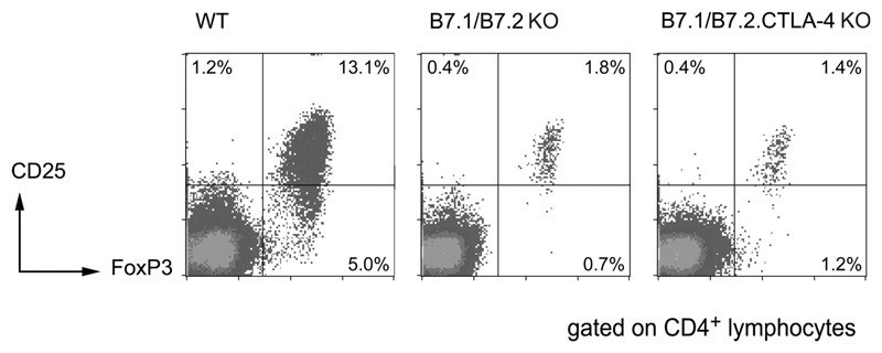
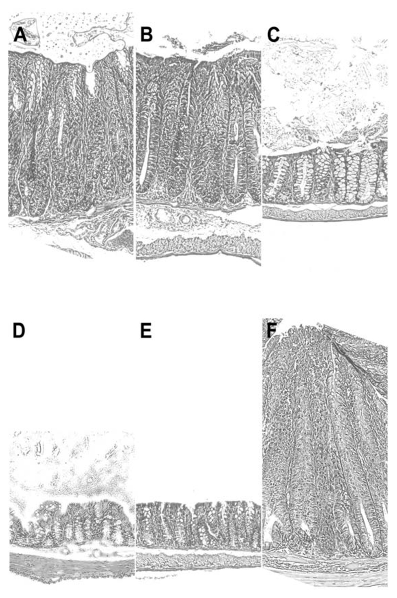
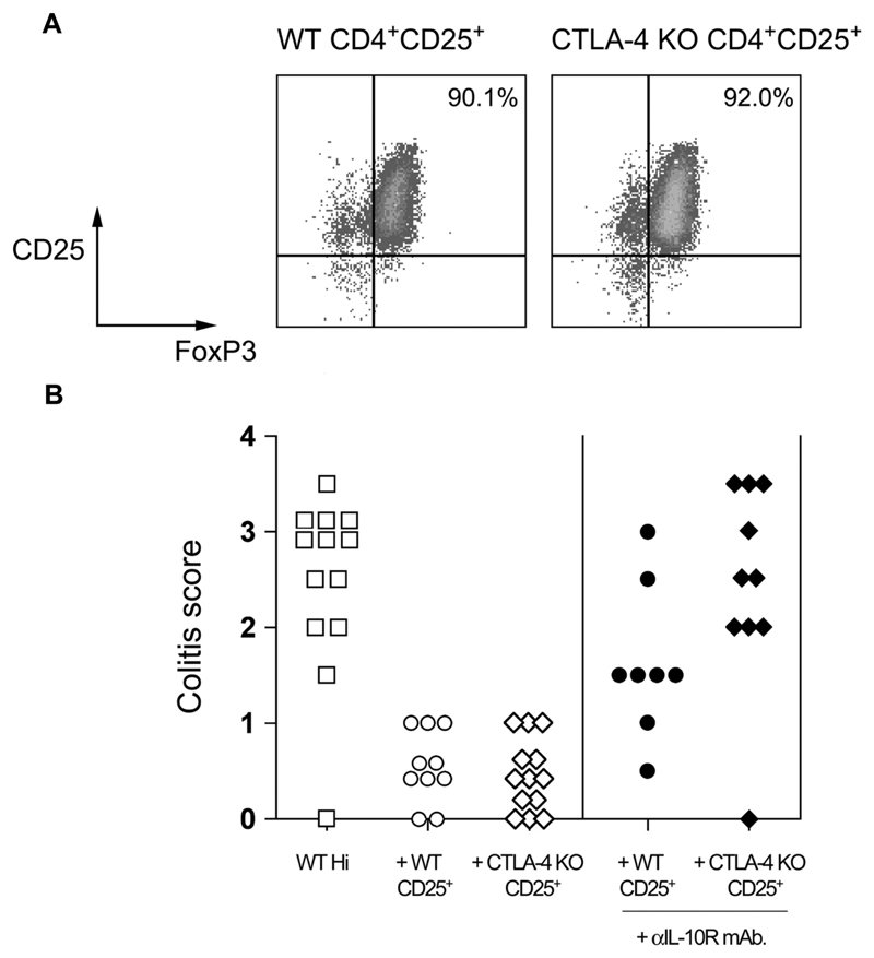
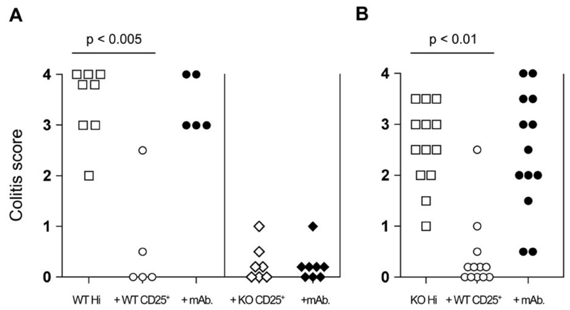
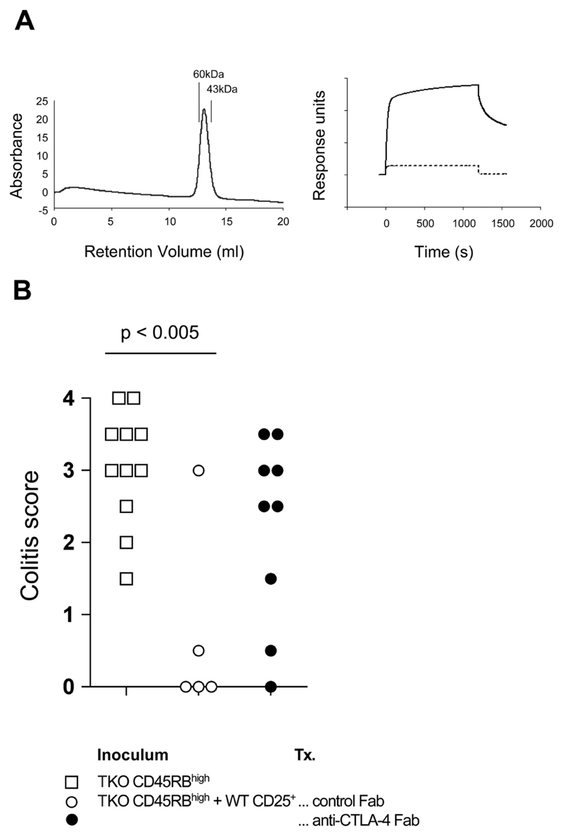
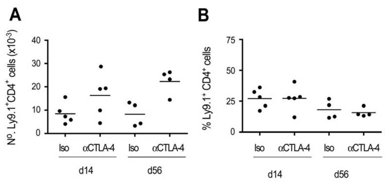
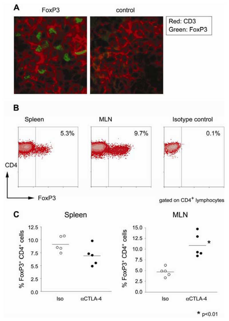
Naturally occurring CD4+ regulatory T cells (T(R)) that express CD25 and the transcription factor FoxP3 play a key role in immune homeostasis, preventing immune pathological responses to self and foreign Ags. CTLA-4 is expressed by a high percentage of these cells, and is often considered as a marker for T(R) in experimental and clinical analysis. However, it has not yet been proven that CTLA-4 has a direct role in T(R) function. In this study, using a T cell-mediated colitis model, we demonstrate that anti-CTLA-4 mAb treatment inhibits T(R) function in vivo via direct effects on CTLA-4-expressing T(R), and not via hyperactivation of colitogenic effector T cells. Although anti-CTLA-4 mAb treatment completely inhibits T(R) function, it does not reduce T(R) numbers or their homing to the GALT, suggesting the Ab mediates its function by blockade of a signal required for T(R) activity. In contrast to the striking effect of the Ab, CTLA-4-deficient mice can produce functional T(R), suggesting that under some circumstances other immune regulatory mechanisms, including the production of IL-10, are able to compensate for the loss of the CTLA-4-mediated pathway. This study provides direct evidence that CTLA-4 has a specific, nonredundant role in the function of normal T(R). This role has to be taken into account when targeting CTLA-4 for therapeutic purposes, as such a strategy will not only boost effector T cell responses, but might also break T(R)-mediated self-tolerance.


Figures

Figure 1
Figure 1. CD4+CD25+FoxP3+ cells are present in both B7-1/B7-2 KO and B7-1/B7-2/CTLA-4 KO mice.
Unfractionated splenocytes from WT, B7-1/B7-2 KO, and B7-1/B7-2/CTLA-4 KO mice were analysed by flow cytometry for the expression of CD4, CD25, and FoxP3. Mice were analysed at 6–8 wk of age. Representative plots show log10 fluorescence and are gated on CD4+ lymphocytes.
Figure 2
Figure 2. Effects of CTLA-4 on development and prevention of colitis.
The figure shows representative photomicrographs of the distal colon of C.B-17 scid mice following transfer of CD4+ T cells. CD4+CD45RBhigh cells isolated from either WT (A) or B7-1/B7-2/CTLA-4 KO mice (B) were able to induce colitis in C.B-17 scid recipients. Cotransfer of WT CD4+CD25+ cells was able to prevent colitis induced by either WT (C) or B7-1/B7-2/CTLA-4 KO CD4+CD45RBhigh cells (D). However, CD4+CD25+ cells from B7-1/B7-2/CTLA-4 KO mice could inhibit colitis induced by WT CD4+ CD45RBhigh cells (E) but not that induced by the equivalent population from B7-1/B7-2/CTLA-4 KO mice (F). Original magnification: ×250 (H&E).
Figure 3
Figure 3. CTLA-4 KO CD4+CD25+ T cells express FoxP3 and retain the ability to prevent colitis.
(A) WT and CTLA-4 KO TR were isolated from CTLA-4 KO mixed bone marrow chimeras by sorting for Ly9.1+CD4+CD25+ (CTLA-4 KO) and Ly9.1CD4+CD25+ (CTLA-4-sufficient) populations as described above. The resulting populations were stained for expression of FoxP3 and analyzed by flow cytometry. Plots are gated on CD4+ lymphocytes, and percentages refer to the percentage of CD25+ FoxP3+ cells. (B) BALB/c RAG KO mice received WT CD4+CD45RBhigh cells alone or in combination with WT or CTLA-4 KO CD4+CD25+ cells. Some mice also received anti-IL10R mAb. Mice were sacrificed 8–10 wk after transfer and colons taken for histological analysis. Data are pooled from two independent experiments. In the presence of anti-IL-10R mAb, WT CD4+CD25+ TR mediated significant protection from colitis (p < 0.05), however, CTLA-4 KO CD4+CD25+ TR provided no significant protection from colitis (p > 0.05).
Figure 4
Figure 4. Anti-CTLA-4 acts to inhibit CD4+CD25+ TR activity by interacting with CTLA-4 expressed by CD4+CD25+ cells.
(A) C.B-17.scid mice received WT CD4+CD45RBhigh cells alone or in combination with either WT or B7-1/B7-2/CTLA-4 KO (triple KO (TKO)) CD4+CD25+ cells. In addition, some mice also received anti-CTLA-4 mAb. (B) In similar experiments, C.B-17.scid mice received B7-1/B7-2/CTLA-4 KO CD4+CD45RBhigh cells alone or in combination with WT CD4+CD25+ cells. Again some mice also received anti-CTLA-4 mAb. Mice were sacrificed 6–8 wk after transfer and colons taken for histological analysis. Data show colitis scores for individual mice taken from two to three independent experiments.
Figure 5
Figure 5. Administration of anti-CTLA-4 Fab is sufficient to abrogate CD4+CD25+ T cell-mediated control of colitis.
(A) Biochemical analyses of anti-CTLA-4 Fab. Left: size-exclusion chromatography indicates that the purified anti-CTLA-4 Fab exists primarily as a monomer. Purified Fab was passed over a Superdex 200 column and eluted as a single peak consistent with a monomer of ~50 kDa. The column was calibrated with molecular mass standards as indicated. In addition, anti-CTLA-4 Fab retained the ability to bind a CTLA-4.Ig fusion protein as measured by surface plasmon resonance (right). The plot shows binding of anti-CTLA-4 Fab to immobilized CTLA-4.Ig as compared with a control reference surface (dotted line). A control Fab failed to bind the immobilized CTLA-4.Ig, giving a trace identical with the reference surface (data not shown). (B) C.B-17 scid mice received B7-1/B7-2/CTLA-4 KO (triple KO (TKO)) CD4+CD45RBhigh cells alone or in combination with CD4+CD25+ cells. Mice also received either anti-CTLA-4 Fab or a control hamster IgG Fab. Mice were sacrificed 6–8 wk after transfer and colons taken for histological analysis. Data show colitis scores for individual mice taken from two independent experiments. In the presence of anti-CTLA-4 Fab, CD4+CD25+ cells failed to confer significant protection from colitis induced by B7-1/B7-2/CTLA-4 KO CD4+CD45RBhigh cells.
Figure 6
Figure 6. Anti-CTLA-4 mAb does not inhibit CD4+CD25+ TR accumulation in vivo.
RAG2 KO mice received BALB/c.C57B10D2.Ly9.2 congenic CD4+CD45RBhigh cells and BALB/c (Ly9.1+) CD4+CD25+ cells together with anti-CTLA-4 or control mAb. Mice were sacrificed and the number and proportion of CD4+Ly9.1+ cells determined at the time points indicated. Data show absolute numbers of CD4+Ly9.1+ cells (A) and Ly9.1+ cells as a proportion of total CD4+ cells (B) in the spleens of individual mice, and are representative of three independent experiments.
Figure 7
Figure 7. CD4+FoxP3+ T cells accumulate in the GALT of diseased mice receiving anti-CTLA-4 mAb.
RAG2 KO mice received CD4+CD45RBhigh and CD4+CD25+ T cells together with anti-CTLA-4 mAb. Eight weeks later, mice were sacrificed and tissues taken for analysis. (A) Sections from mesenteric lymph nodes were stained for CD3 (red) and FoxP3 (green), or CD3 and a control rabbit Ig. Images are representative of the analysis of four mice from two independent experiments. Original magnification: ×630. (B) Representative analysis of FoxP3 expression by transferred CD4+ T cells from anti-CTLA-4-treated mice. Single-cell suspensions from spleen and mesenteric lymph nodes were stained for flow cytometric analysis. Plots show log10 fluorescence and are gated on CD3+CD4+ lymphocytes. (C) Proportion of CD4+ T cells expressing FoxP3 in the spleen and mesenteric lymph nodes of anti-CTLA-4 or control mice. Each point represents a single mouse analyzed by flow cytometry as in (B).

References

    1. Shevach EM. CD4+CD25+ suppressor T cells: more questions than answers. Nat Rev Immunol. 2002;2:389–400. - PubMed
    1. Belkaid Y, Rouse BT. Natural regulatory T cells in infectious disease. Nat Immunol. 2005;6:353–360. - PubMed
    1. Pirzer U, Schonhaar A, Fleischer B, Hermann E, Meyer zum Buschenfelde KH. Reactivity of infiltrating T lymphocytes with microbial antigens in Crohn’s disease. Lancet. 1991;338:1238–1239. - PubMed
    1. Duchmann R, Kaiser I, Hermann E, Mayet W, Ewe K, Meyer zum Buschenfelde KH. Tolerance exists towards resident intestinal flora but is broken in active inflammatory bowel disease (IBD) Clin Exp Immunol. 1995;102:448–455. - PMC - PubMed
    1. Coombes JL, Robinson NJ, Maloy KJ, Uhlig HH, Powrie F. Regulatory T cells and intestinal homeostasis. Immunol Rev. 2005;204:184–194. - PubMed

Publication types

MeSH terms