. 2006 Sep 26;103(39):14337-42.
doi: 10.1073/pnas.0606603103. Epub 2006 Sep 18.

Crystal structure of 12-oxophytodienoate reductase 3 from tomato: self-inhibition by dimerization

Affiliations

Crystal structure of 12-oxophytodienoate reductase 3 from tomato: self-inhibition by dimerization

Constanze Breithaupt et al. Proc Natl Acad Sci U S A. .

Abstract

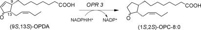
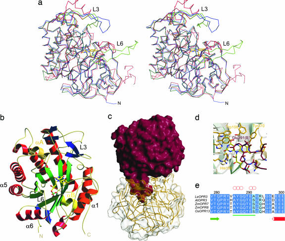
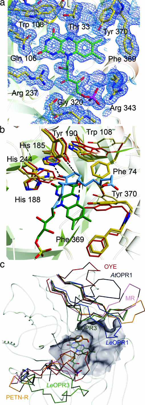
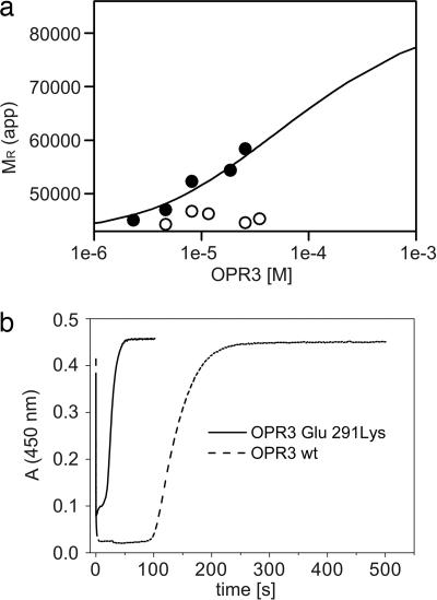
12-Oxophytodienoate reductase (OPR) 3, a homologue of old yellow enzyme (OYE), catalyzes the reduction of 9S,13S-12-oxophytodienoate to the corresponding cyclopentanone, which is subsequently converted to the plant hormone jasmonic acid (JA). JA and JA derivatives, as well as 12-oxophytodienoate and related cyclopentenones, are known to regulate gene expression in plant development and defense. Together with other oxygenated fatty acid derivatives, they form the oxylipin signature in plants, which resembles the pool of prostaglandins in animals. Here, we report the crystal structure of OPR3 from tomato and of two OPR3 mutants. Although the catalytic residues of OPR3 and related OYEs are highly conserved, several characteristic differences can be discerned in the substrate-binding regions, explaining the remarkable substrate stereoselectivity of OPR isozymes. Interestingly, OPR3 crystallized as an extraordinary self-inhibited dimer. Mutagenesis studies and biochemical analysis confirmed a weak dimerization of OPR3 in vitro, which correlated with a loss of enzymatic activity. Based on structural data of OPR3, a putative mechanism for a strong and reversible dimerization of OPR3 in vivo that involves phosphorylation of OPR3 is suggested. This mechanism could contribute to the shaping of the oxylipin signature, which is critical for fine-tuning gene expression in plants.


Conflict of interest statement

The authors declare no conflict of interest.

Figures

Fig. 1.
Fig. 1.
OPR3 catalyzes the reduction of 9S,13S-OPDA to 1S,2S-3-oxo- 2(2′[Z]-pentenyl)-cyclopentane-1-octanoate (OPC-8:0).
Fig. 2.
Fig. 2.
Overall structure of OPR3 from L. esculentum. (a) Stereoview representation of the superimposed Cα traces of OPR3 (green), OPR1 (blue), and OYE (red). (b) Ribbon representation of one OPR3 monomer. The FMN cofactor is depicted as a stick model. (c) Overall structure of the OPR3 dimer. The finger-like loop L6 of protomer A (red) is bound into the substrate-binding pocket of protomer B (yellow) and vice versa. (d) Detailed view of the active site of OPR3 (yellow) blocked by amino acids of loop L6 of the partner protomer (red). (e) Multiple sequence alignment of the insertion (underlined in green) in loop L6 of OPR3-like enzymes. Red circles indicate amino acids that form hydrogen bonds to active-site amino acids.
Fig. 3.
Fig. 3.
Active site of OPR3. (a) FMN-binding site. FMN (green) and amino acids that contact the FMN (yellow) are shown as stick models. The 2FoFc electron density map is calculated at 1.5 Å and contoured at 1.0σ. (b) Superposition of the substrate-binding pockets of OPR3 (yellow), OPR1 (red), and OYE (beige). In addition, the FMN of OPR3 (green) and the OPR1 substrate 9R,13R-OPDA (blue) are shown to visualize interactions that are essential for catalysis. (c) Cα backbone of OPR3 and superimposed L3 and L6 loops of members of the OYE family. LeOPR3, light green; AtOPR3, dark green, LeOPR1, blue; AtOPR1, dark blue; OYE, red; morphinone reductase (MR), magenta; pentaerythritol tetranitrate reductase (PETN-R), yellow. In addition, the surface of the substrate-binding pocket of OPR3 and p-hydroxy benzaldehyde complexed to OYE are shown.
Fig. 4.
Fig. 4.
Biochemical analysis of the OPR3 dimer. (a) Sedimentation equilibrium of WT OPR3 (filled circles) and the Glu-291–Lys mutant (open circles) was analyzed at a protein concentration of 1–34 μM in the presence of 50 μM FMN. For WT OPR3, the apparent molecular masses were fitted to a monomer dimer equilibrium, yielding a dissociation constant (Kd) of 30 μM. (b) Enzyme-monitored turnover. WT OPR3 (solid line) and Glu-291–Lys OPR3 (dashed line) at 86 μM were mixed in the stopped-flow instrument with 1.4 mM NADPH and 1.5 mM trans-hex-2-enal (100 mM potassium phosphate, pH 7; 25°C). In both cases, rapid reduction by NADPH was observed (time = 0 s), followed by a steady-state phase. After exhaustion of NADPH, the enzyme returned to its oxidized state, as indicated by the recovery of absorbance at 450 nm.
Fig. 5.
Fig. 5.
Central section of the dimer interface of protomer A (red; amino acids are marked with an asterisk) and protomer B (yellow) of OPR3. In the crystal structure, a sulfate ion mediates various intermolecular contacts. The position of the sulfate ion would fit very well with that of the phosphate group of a phosphorylated Tyr-364 (blue).

References

    1. Warburg O, Christian W. Biochem Z. 1935;266:377–411.
    1. Massey V. Biochem Soc Trans. 2000;28:283–296. - PubMed
    1. Williams RE, Bruce NC. Microbiology. 2002;148:1607–1614. - PubMed
    1. Karplus PA, Fox KM, Massey V. FASEB J. 1995;9:1518–1526. - PubMed
    1. Stintzi A, Browse J. Proc Natl Acad Sci USA. 2000;97:10625–10630. - PMC - PubMed

Publication types

MeSH terms

Substances

LinkOut - more resources