. 2006 Sep 26;103(39):14537-41.
doi: 10.1073/pnas.0604144103. Epub 2006 Sep 18.

Ex vivo pretreatment of bone marrow mononuclear cells with endothelial NO synthase enhancer AVE9488 enhances their functional activity for cell therapy

Affiliations

Ex vivo pretreatment of bone marrow mononuclear cells with endothelial NO synthase enhancer AVE9488 enhances their functional activity for cell therapy

Ken-ichiro Sasaki et al. Proc Natl Acad Sci U S A. .

Abstract

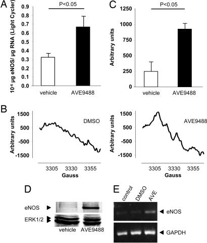
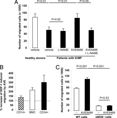
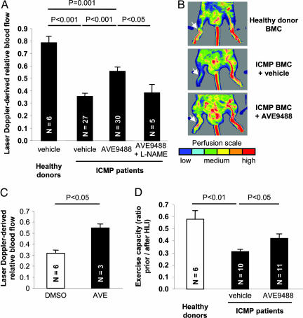
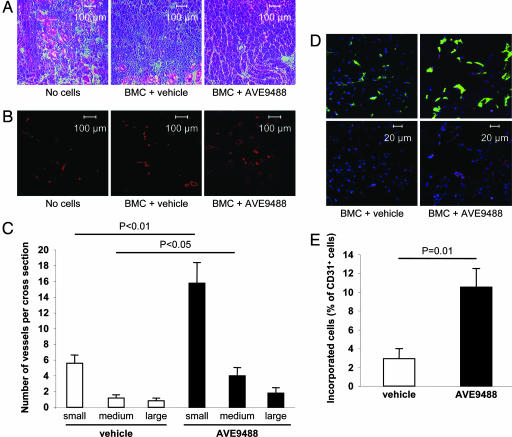
Bone marrow mononuclear cells (BMC) from patients with ischemic cardiomyopathy (ICMP) show a reduced neovascularization capacity in vivo. NO plays an important role in neovascularization, and NO bioavailability is typically reduced in patients with ICMP. We investigated whether the impaired neovascularization capacity of ICMP patient-derived progenitor cells can be restored by pretreatment with the novel endothelial NO synthase (eNOS) transcription enhancer AVE9488 (AVE). Ex vivo pretreatment of BMC from patients with ICMP with AVE significantly increased eNOS mRNA expression by 2.1-fold (P < 0.05) and eNOS activity as assessed by ESR by >3-fold (P < 0.05). The increased eNOS expression was associated with an enhanced migratory capacity in vitro (P < 0.01) and improved neovascularization capacity of the infused BMC in an ischemic hind limb model in vivo (P < 0.001). The improvement in ischemic limb perfusion after infusion of AVE-pretreated BMC resulted in an increase in swimming time (P < 0.05). The enhancement of limb perfusion by AVE-treated BMC was abrogated by ex vivo pretreatment with the eNOS inhibitor N(G)-nitro-l-arginine methyl ester. Consistently, AVE showed no effect on the impaired migratory capacity of BMC derived from eNOS-deficient mice, documenting the specific involvement of NO. The reduced neovascularization capacity of BMC from patients with ICMP may limit their therapeutic potential in cell therapy studies. Here, we show that pharmacological enhancement of eNOS expression with AVE at least partially reverses the impaired functional activity of BMC from ICMP patients, highlighting the critical role of NO for progenitor cell function.


Conflict of interest statement

Conflict of interest statement: S.D., A.M., and C.H. have filed for a patent for the use of eNOS transcription enhancers in cell therapy of ischemic heart diseases (U.S. application 20050101599).

Figures

Fig. 1.
Fig. 1.
Effect of AVE on eNOS expression. (A) Expression of eNOS RNA, protein, and eNOS-derived NO is augmented in the presence of the eNOS transcription enhancer AVE. eNOS mRNA expression was detected by quantitative real-time RT-PCR of 100 ng of RNA by using a light cycler instrument in BMC derived from patients with ICMP pretreated with vehicle or AVE (5 μM) for 18 h (n = 3). (B and C) NO production of LinCD105+ cells was determined by ESR. (B) Representative recordings from n = 3 experiments are depicted. (C) Quantitative data are presented. (D) eNOS Western blot analysis of cells treated with either vehicle or AVE. A representative blot is shown. ERK1/2 was used as a loading control. (E) eNOS expression was measured by RT-PCR in CD34+ cells after treatment with 5 μM AVE for 24 h.
Fig. 2.
Fig. 2.
Effect of AVE on migration. (A) Shown are the numbers of migrated BMC toward the chemoattractant factor SDF-1 derived from healthy donors (n = 12) and patients with ICMP that were pretreated for 18 h with either vehicle (n = 11), l-NAME (n = 11), AVE (n = 11), or AVE plus l-NAME (n = 6). (B) Increase in SDF-1-mediated migration by AVE-pretreatment (5 μM, 24 h) in CD14+ cells, BMC, and CD34+ cells isolated from healthy volunteers (n = 3–4) is shown. (C) The number of BMC from WT and eNOS knockout (eNOS−/−) mice that migrated in response to SDF-1 to the lower chamber of a modified Boyden chamber assay after pretreatment for 18 h with either vehicle or AVE (n ≥ 8 per group) is shown.
Fig. 3.
Fig. 3.
Effect of AVE on recovery after ischemia. (A) Recovery of limb perfusion after hind limb ischemia as assessed by laser Doppler blood flow analysis on day 14 after i.v. infusion of BMC derived from either healthy donors or nine patients with ICMP into nude mice. BMC from ICMP patients were pretreated with vehicle, AVE, or AVE plus l-NAME for 18 h. The number of mice in each group is indicated in the bars. (B) Representative laser Doppler images for each group are depicted. Arrows indicate ischemic limbs. Perfusion signals are subdivided into six different intervals, each displayed as a separate color. Low or no perfusion is displayed as dark blue; highest perfusion interval is displayed as red. (C) Recovery of limb perfusion by i.v. infusion of circulating blood-derived EPC isolated from patients after pretreatment with AVE for 18 h compared with controls. (D) Exercise capacity expressed as the swimming time ratio (swimming time on day 14 after hind limb ischemia/swimming time before induction of hind limb ischemia) in a murine model of hind limb ischemia. Mice were either untreated or received i.v. infusions of BMC from healthy donors or patients with ICMP.
Fig. 4.
Fig. 4.
Effect of AVE on morphology, capillary density, and incorporation. (A) Histological morphology of the ischemic muscles was assessed in H&E-stained sections. (B) Conductance vessels were identified by staining for smooth muscle α-actin with a Cy3-labeled mouse mAb (red color). (C) The number of small (<50 μm), medium (50–100 μm), and large (>100 μm) vessels was counted separately (n = 5 mice per group). (D) Incorporation of human BMC (stained for HLA-ABC, red) into vessels (endothelial marker CD31, green) after pretreatment with vehicle or AVE. Sytox was used for nuclear staining (blue). Representative images are shown. (Upper) Merged pictures. (Lower) Staining for human HLA and nuclei. (E) Double positive cells were quantified from 10 sections provided by five mice per group.

References

    1. Kocher AA, Schuster MD, Szabolcs MJ, Takuma S, Burkhoff D, Wang J, Homma S, Edwards NM, Itescu S. Nat Med. 2001;7:430–436. - PubMed
    1. Orlic D, Kajstura J, Chimenti S, Jakoniuk I, Anderson SM, Li B, Pickel J, McKay R, Nadal-Ginard B, Bodine DM, et al. Nature. 2001;410:701–705. - PubMed
    1. Asahara T, Murohara T, Sullivan A, Silver M, van der Zee R, Li T, Witzenbichler B, Schatteman G, Isner JM. Science. 1997;275:964–967. - PubMed
    1. Assmus B, Schachinger V, Teupe C, Britten M, Lehmann R, Dobert N, Grunwald F, Aicher A, Urbich C, Martin H, et al. Circulation. 2002;106:3009–3017. - PubMed
    1. Perin EC, Dohmann HF, Borojevic R, Silva SA, Sousa AL, Mesquita CT, Rossi MI, Carvalho AC, Dutra HS, Dohmann HJ, et al. Circulation. 2003;107:2294–2302. - PubMed

Publication types

MeSH terms

LinkOut - more resources