Comparative Study
. 2006 Sep 26;103(39):14385-9.
doi: 10.1073/pnas.0603529103. Epub 2006 Sep 18.

Differences in vertebrate microRNA expression

Affiliations
Comparative Study

Differences in vertebrate microRNA expression

Brandon Ason et al. Proc Natl Acad Sci U S A. .

Abstract

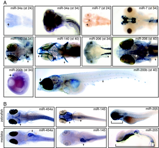
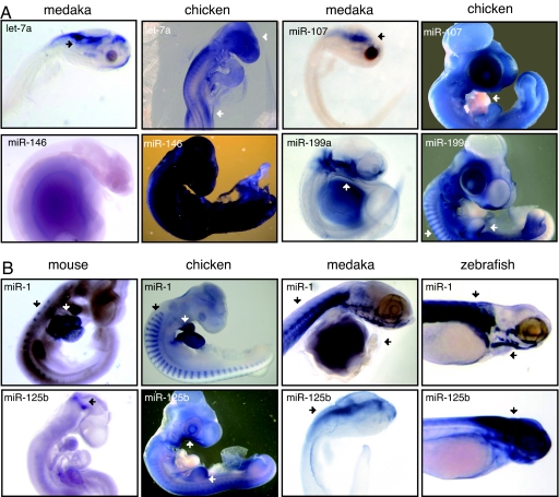
MicroRNAs (miRNAs) attenuate gene expression by means of translational inhibition and mRNA degradation. They are abundant, highly conserved, and predicted to regulate a large number of transcripts. Several hundred miRNA classes are known, and many are associated with cell proliferation and differentiation. Many exhibit tissue-specific expression, which aids in evaluating their functions, and it has been assumed that their high level of sequence conservation implies a high level of expression conservation. A limited amount of data supports this, although discrepancies do exist. By comparing the expression of approximately 100 miRNAs in medaka and chicken with existing data for zebrafish and mouse, we conclude that the timing and location of miRNA expression is not strictly conserved. In some instances, differences in expression are associated with changes in miRNA copy number, genomic context, or both between species. Variation in miRNA expression is more pronounced the greater the differences in physiology, and it is enticing to speculate that changes in miRNA expression may play a role in shaping the physiological differences produced during animal development.


Conflict of interest statement

The authors declare no conflict of interest.

Figures

Fig. 1.
Fig. 1.
MiRNA expression in medaka. (A) MiRNAs are expressed late in medaka development. Representative examples are shown. Brain tissue is clearly visible by stage 24 (see miR-34a and miR-7, arrows), yet peak expression occurs much later, stage 34. MiRNA expression is further delayed in tissues associated with feeding such as the jaw and head muscles, as well as sense organs such as the lateral line and taste buds. Representative examples of cartilage (miR-140), muscle (miR-206), and sensory epithelia (miR 200b) specific expression are shown. In each case, peak expression occurs later in these tissues (stage 40, arrows) compared with expression of the same miRNA in related tissues (stage 34, arrows). (B) Several miRNAs exhibit spatial expression differences between medaka and zebrafish. Representative examples are shown. MiR-454a is expressed in the gut (arrow) in medaka and in the dorsal midbrain (arrow) in zebrafish. MiR-145 is observed in zebrafish pharyngeal arches (arrow) and fins with weaker expression observed in the gut and gall bladder (arrow). In medaka, expression is most pronounced in the heart and gut (arrow), with weak expression observed late in the pharyngeal arches and head skeleton. MiR-205 is expressed in the epidermis in both medaka and zebrafish. Expression extends through the entire length of the lateral surface (bracket) in medaka (stage 34) but strong expression is limited to the epidermis surrounding the pharyngeal arches in zebrafish (bracket). During later developmental stages, strong expression in medaka is limited to the pharyngeal arches, mimicking observed expression in zebrafish (data not shown).
Fig. 2.
Fig. 2.
Comparison of miRNA expression. (A) Regional differences in miRNA expression are observed between chicken and medaka. For let-7a, brain-specific expression is seen for both medaka and chicken (arrows). In chicken, additional expression is also observed in the developing limb buds (arrow). MiR-199a is expressed in the epithelia surrounding the pharyngeal arches in medaka (arrows), whereas expression is observed in head, limb, and body mesoderm in chicken (arrows). In chicken, miR-107 is expressed in all tissues, except the heart (arrow). In medaka, miR-107 expression is brain-specific (arrow). MiR-146 is specifically expressed in the gut late in medaka development (stage 40), exhibiting no expression in earlier stages (shown). In chicken, miR-146 is expressed in the epidermis early in development, leading to ubiquitous expression in later developmental stages. (B) Comparison of miRNA expression among mouse, chicken, medaka, and zebrafish. Representative examples of differences in expression are shown. MiR-1 is expressed in skeletal muscle in both medaka and zebrafish (arrows). Similar expression is observed in the progenitor to muscle, or somites, in both chicken and mouse (arrows) and is additionally expressed in the heart (arrows). MiR-125b is expressed in the brain and spinal cord in both medaka and zebrafish (arrows). It is ubiquitously expressed in chicken, with stronger expression in the pharyngeal arches and limb buds (arrows). In mouse, expression is observed at the midbrain–hindbrain boundary (arrow).

References

    1. Bartel DP. Cell. 2004;116:281–297. - PubMed
    1. Alvarez-Garcia I, Miska EA. Development (Cambridge, UK) 2005;132:4653–4662. - PubMed
    1. Plasterk RH. Cell. 2006;124:877–881. - PubMed
    1. He L, Thomson JM, Hemann MT, Hernando-Monge E, Mu D, Goodson S, Powers S, Cordon-Cardo C, Lowe SW, Hannon GJ, Hammond SM. Nature. 2005;435:828–833. - PMC - PubMed
    1. Lu J, Getz G, Miska EA, Alvarez-Saavedra E, Lamb J, Peck D, Sweet-Cordero A, Ebert BL, Mak RH, Ferrando AA, et al. Nature. 2005;435:834–838. - PubMed

Publication types

LinkOut - more resources