. 2006 Oct 1;20(19):2673-86.
doi: 10.1101/gad.1451806. Epub 2006 Sep 18.

A cancer cell metalloprotease triad regulates the basement membrane transmigration program

Affiliations

A cancer cell metalloprotease triad regulates the basement membrane transmigration program

Kevin Hotary et al. Genes Dev. .

Erratum in

  • Genes Dev. 2007 May 1;21(9):1139

Abstract

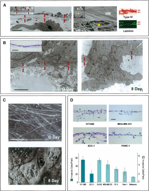
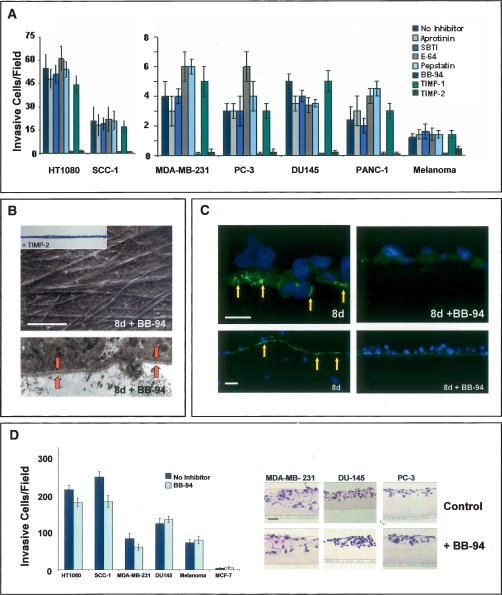
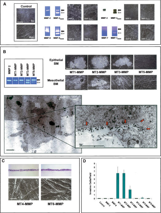
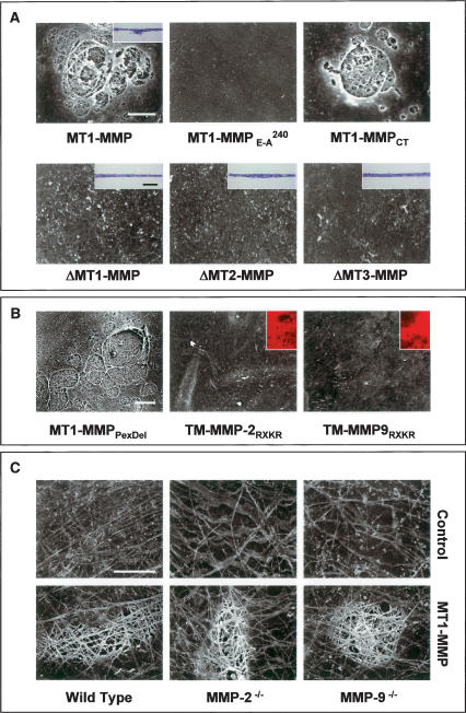
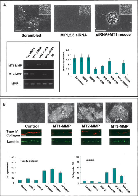
Carcinoma cells initiate the metastatic cascade by inserting invasive pseudopodia through breaches in the basement membrane (BM), a specialized barrier of cross-linked, extracellular matrix macromolecules that underlies epithelial cells and ensheaths blood vessels. While BM invasion is the sine qua non of the malignant phenotype, the molecular programs that underlie this process remain undefined. To identify genes that direct BM remodeling and transmigration, we coupled high-resolution electron microscopy with an ex vivo model of invasion that phenocopies the major steps observed during the transition of carcinoma in situ to frank malignancy. Herein, a triad of membrane-anchored proteases, termed membrane type-1, type-2, and type-3 metalloproteinases, are identified as the triggering agents that independently confer cancer cells with the ability to proteolytically efface the BM scaffolding, initiate the assembly of invasive pseudopodia, and propagate transmigration. These studies characterize the first series of gene products capable of orchestrating the entire BM remodeling program that distinguishes the carcinomatous phenotype.


Figures

Figure 1.
Figure 1.
BM remodeling and transmigration by cancer cells. (A, left) Mesothelial BM (rat peritoneum) shown by TEM (red arrows demarcate BM). (Right) SEM (BM) highlights the sheet-like structure of the BM overlaying the stromal matrix (indicated by an asterisk). Immunostaining for type IV collagen and laminin (horizontal arrows) reveals the two BM layers (i.e., the upper and lower surfaces of the peritoneum are lined by BMs). Insets show the epithelial BM deposited by MDCK cells as visualized by TEM and SEM. Bars for TEM, SEM, and light micrographs are 1, 20, and 100 μm, respectively. (B) MDA-MB-231 cells cultured atop the peritoneal BM at 0 and 8 d as assessed by TEM. BM (highlighted by red arrows) is breached by carcinoma cells after an 8-d culture period. Inset shows an H&E section of carcinoma cells at 0 d. Bars for TEM and light micrographs are 1 and 200 μm, respectively. (C) SEM of peritoneal BM stripped of overlying MDA-MB-231 cancer cells immediately after plating (0 d), or after an 8-d culture period (8 d). Bar, 10 μm. (D) Representative H&E cross-sections of the peritoneum after an 8-d culture period. The number of invasive cells per field are quantified in H&E-stained cross-sections with error bars indicating the standard error of mean of five or more experiments.
Figure 2.
Figure 2.
MMP-dependent BM invasion. (A) Cancer cells were cultured atop epithelial BMs for 8 d in the absence or presence of inhibitors directed against serine proteinases (aprotinin, soybean trypsin inhibitor; SBTI), cysteine proteinases (E-64), aspartyl proteinases (pepstatin) or MMPs (BB-94, TIMP-1, TIMP-2). Invasion is shown as the mean number of invasive cells per field ± standard error of mean of five or more experiments. (B) SEM and TEM analyses of peritoneal BM stripped of overlying MDA-MB-231 cells following an 8-d culture period in the absence or presence of BB-94. (Bottom) The intact BM (which also completely surrounds the cellular projection from the basal surface) is bracketed by the red arrows in the TEM micrograph. Similar results were obtained with all of the cancer cell lines studied (data not shown). The inset shows the absence of invasion (as assessed in an H&E-stained cross-section) when cancer cells are cultured in the presence of TIMP-2. Bar, 50 μm. (C) Degraded type IV collagen (green) is detected in peritoneal BMs breached by invasive MDA-MB-231 cells following an 8-d culture period (yellow arrows; the top and bottom images highlight the pattern of type IV collagen degradation at high and low magnification, respectively). In the presence of BB-94, MDAMB-231 cells neither degrade nor invade the underlying BM following an 8-d culture period. Cells are stained with DAPI (blue). Bars for SEM and light micrographs are 50 and 100 μm, respectively. (D) Tumor cell invasion through Matrigel-coated filters was quantified following an 48-h culture period in the absence or presence of BB-94. The number of invasive cells per field is expressed as the mean ± standard error of mean (n = 4). H&E-stained cross-sections of tumor cell invasion through thick gels of Matrigel in the absence or presence of BB-94 following a 4-d culture period are shown to the right.
Figure 3.
Figure 3.
MT-MMP-mediated BM transmigration. (A) Control COS cells or COS cells transiently expressing either wild-type or RXKR (active) forms of MMP-2, MMP-9, MMP-3, or MMP-7 were cultured on mesothelial or epithelial BMs for 5 d, detergent-lysed, and visualized by SEM in representative samples (identical results were obtained with either BM type). Black arrows indicate proMMPs and white arrows indicate processed MMPs as determined by zymography (MMP-2, MMP-7, and MMP-9) or Western blot analysis (MMP-3). (B) Following culture of transfected cells atop BMs, SEM analysis shows discrete fields of perforations in epithelial or mesothelial BMs by MT1-MMP, MT2-MMP, or MT3-MMP, but not MT5-MMP-expressing COS cells. Zymograms show processing of exogenous MMP-2 by COS cells expressing MT1-MMP, MT2-MMP, MT3-MMP, or MT5-MMP (box). TEM shows an MT2-MMP-expressing COS cell inserting an invasive pseudopod through the epithelial BM (red arrows in inset) into the underlying stroma. (C) COS cells expressing MT4-MMP or MT6-MMP were unable to invade or remodel the underlying peritoneal BM as assessed in H&E-stained cross-sections or by SEM. (D) The number of invasive COS cells was determined in H&E-stained cross-sections of epithelial BM cultures. Results are shown as the mean number of invasive cells per field ± standard error of mean (n = 3). Bars for TEM and SEM are 1 and 50 μm, respectively.
Figure 4.
Figure 4.
Structure–function analysis of MT-MMP-mediated BM proteolysis. (A) COS cells were transfected with MT1-MMP; catalytically inactive MT1-MMP (MT1-MMPE →A 240); cytoplasmic tail-deleted MT1-MMP (MT1-MMPCT); or soluble, transmembrane-deleted forms of MT1-MMP, MT2-MMP, or MT3-MMP (ΔMT1-MMP, ΔMT2-MMP, or ΔMT3-MMP) and cultured atop epithelial BMs for a 5-d culture period. Cells were removed and the underlying BM was assessed by SEM. Insets show representative H&E-stained cross-sections. (B) COS cells were transfected with an MT1-MMP construct wherein the hemopexin domain was deleted (MT1-MMPPexDel) or, alternatively, with chimeric forms of either membrane-anchored, active MMP-2 or MMP-9 (TM MMP-2RXKR or TM-MMP-9RXKR, respectively). Following a 5-d culture period, BM structure was assessed by SEM. Insets demonstrate the ability of the membrane-anchored gelatinases to degrade a subjacent bed of fluorescent gelatin. (C) Mesothelial BMs isolated from wild-type, MMP-2−/−, or MMP-9−/− mice were visualized by SEM after a 5-d culture period in the respective null serum with control or MT1-MMP-transfected COS cells. Bars for SEM and light micrographs are 10 and 100 μm, respectively.
Figure 5.
Figure 5.
MT-MMP-dependent control of carcinoma cell BM invasion. (A) MDA-MB-231 cells were electroporated with either a scrambled siRNA or MT1-MMP, MT2-MMP, and MT3-MMP siRNA in combination, and then cotransfected with a control or mouse MT1-MMP expression vector (rescue). After a 5-d culture period atop epithelial BMs, matrices were denuded and examined by SEM. Insets show tumor cell morphology following electroporation as assessed by phase contrast microscopy. MT1-MMP, MT2-MMP, and MT3-MMP siRNAs inhibited expression of the targeted proteases, but had no effect on the expression of nontargeted MMPs as shown by RT–PCR (box). The chart shows the average number of BM perforations quantified in 25 randomly selected fields for each siRNA transfection and rescue (mean ± standard error of mean; n = 3). (B) Epithelial BM structure, as assessed by SEM (with corresponding type IV collagen and laminin immunofluorescence below), following a 5-d culture period with control-, MT1-MMP-, MT2-MMP-, or MT3-MMP-transfected MCF-7 cells. Bar charts depict the percent degraded BM (i.e., loss of visible type IV collagen or laminin fluorescence) quantified in 10 random fields. All bars are shown ± standard error of the mean. Bars for SEM and light micrographs are 10 and 100 μm, respectively.

References

    1. Abrams, G.A., Goodman, S.L., Nealey, P.F., Franco, M., Murphy, C.J. Nanoscale topography of the basement membrane underlying the corneal epithelium of the rhesus macaque. Cell Tissue Res. 2000;299:39–46. - PubMed
    1. Aplin, J.D., Campbell, S., Allen, T.D. The extracellular matrix of human amniotic epithelium: Ultrastructure, composition and deposition. J. Cell Sci. 1985;79:119–136. - PubMed
    1. Baluk, P., Raymond, W.W., Ator, E., Coussens, L.M., McDonald, D.M., Caughey, G.H. Matrix metalloproteinase-2 and -9 expression increases in mycoplasma-infected airways but is not required for microvascular remodeling. Am. J. Physiol. Lung Cell. Mol. Physiol. 2004;287:L307–L317. - PubMed
    1. Brabletz, T., Spaderna, S., Kolb, J., Hlubek, F., Faller, G., Bruns, C.J., Jung, A., Nentwich, J., Duluc, I., Domon-Dell, C., et al. Down-regulation of the homeodomain factor Cdx2 in colorectal cancer by collagen type I: An active role for the tumor environment in malignant tumor progression. Cancer Res. 2004;64:6973–6977. - PubMed
    1. Cao, J., Kozarekar, P., Pavlaki, M., Chiarelli, C., Bahou, W.F., Zucker, S. Distinct roles for the catalytic and hemopexin domains of membrane type 1-matrix metalloproteinase in substrate degradation and cell migration. J. Biol. Chem. 2004;279:14129–14139. - PubMed

Publication types

MeSH terms

Substances

LinkOut - more resources