. 2006 Sep;3(9):e358.
doi: 10.1371/journal.pmed.0030358.

In celiac disease, a subset of autoantibodies against transglutaminase binds toll-like receptor 4 and induces activation of monocytes

Affiliations

In celiac disease, a subset of autoantibodies against transglutaminase binds toll-like receptor 4 and induces activation of monocytes

Giovanna Zanoni et al. PLoS Med. 2006 Sep.

Abstract

Background: Celiac disease is a small intestine inflammatory disorder with multiple organ involvement, sustained by an inappropriate immune response to dietary gluten. Anti-transglutaminase antibodies are a typical serological marker in patients with active disease, and may disappear during a gluten-free diet treatment. Involvement of infectious agents and innate immunity has been suggested but never proven. Molecular mimicry is one of the mechanisms that links infection and autoimmunity.

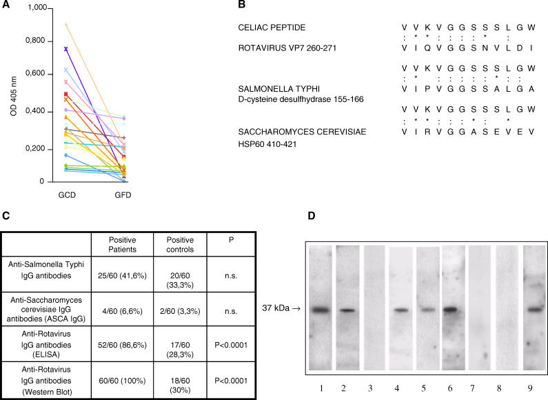
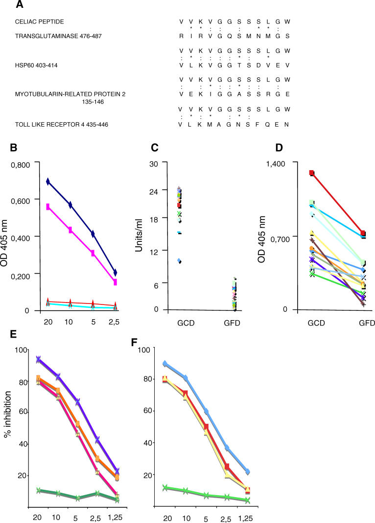
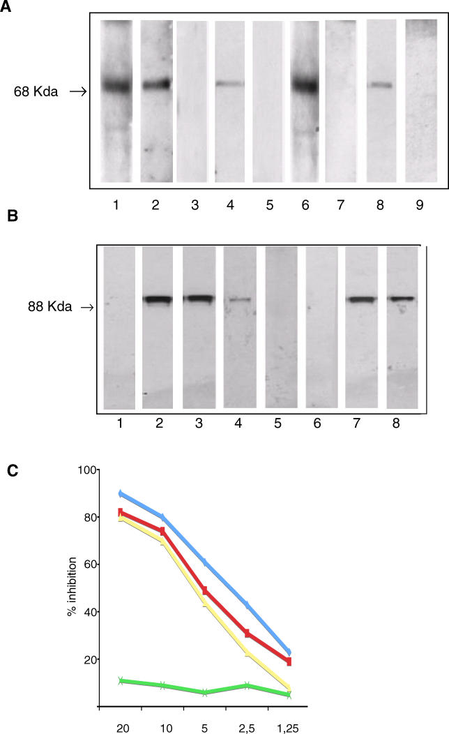
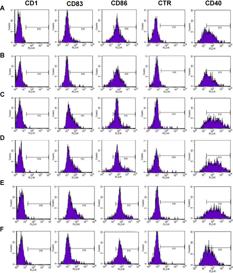
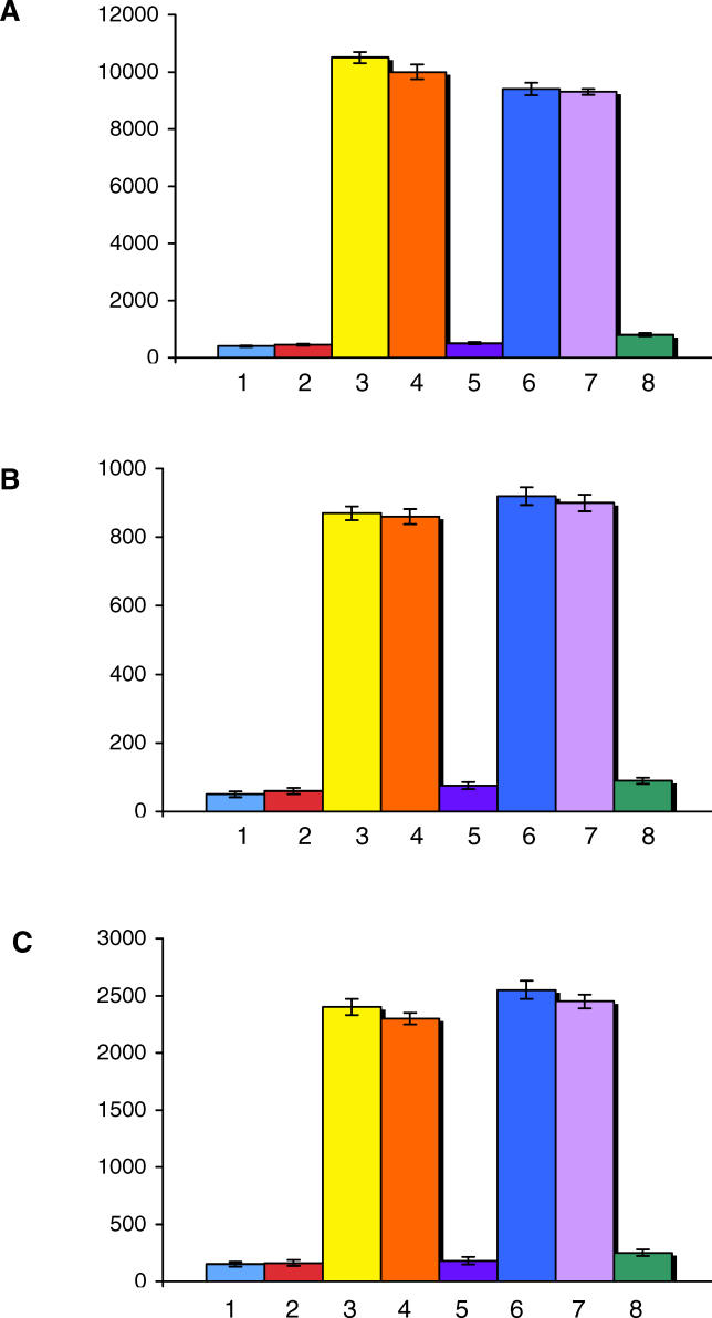
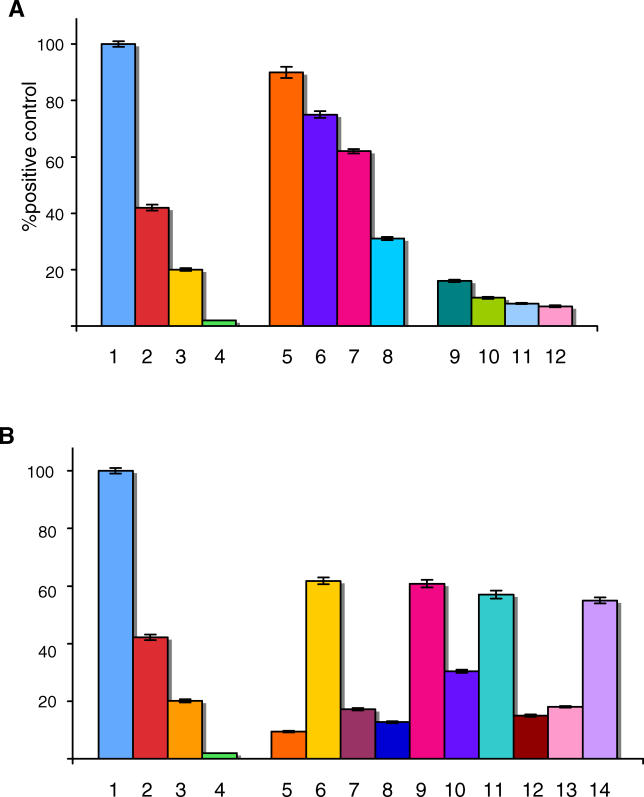
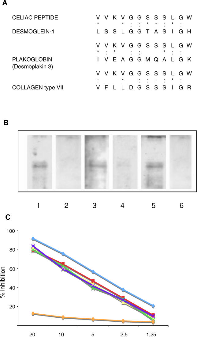
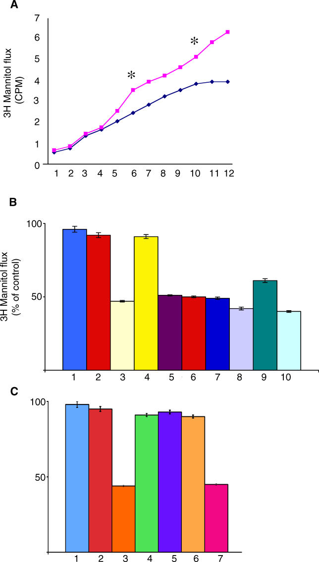
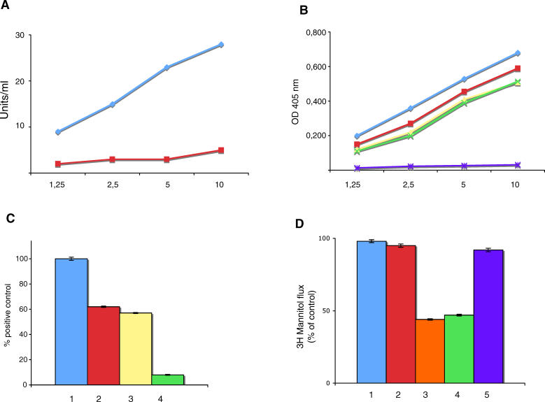
Methods and findings: In our attempt to clarify the pathogenesis of celiac disease, we screened a random peptide library with pooled sera of patients affected by active disease after a pre-screening with the sera of the same patients on a gluten-free diet. We identified a peptide recognized by serum immunoglobulins of patients with active disease, but not by those of patients on a gluten-free diet. This peptide shares homology with the rotavirus major neutralizing protein VP-7 and with the self-antigens tissue transglutaminase, human heat shock protein 60, desmoglein 1, and Toll-like receptor 4. We show that antibodies against the peptide affinity-purified from the sera of patients with active disease recognize the viral product and self-antigens in ELISA and Western blot. These antibodies were able to induce increased epithelial cell permeability evaluated by transepithelial flux of [(3)H] mannitol in the T84 human intestinal epithelial cell line. Finally, the purified antibodies induced monocyte activation upon binding Toll-like receptor 4, evaluated both by surface expression of activation markers and by production of pro-inflammatory cytokines.

Conclusions: Our findings show that in active celiac disease, a subset of anti-transglutaminase IgA antibodies recognize the viral protein VP-7, suggesting a possible involvement of rotavirus infection in the pathogenesis of the disease, through a mechanism of molecular mimicry. Moreover, such antibodies recognize self-antigens and are functionally active, able to increase intestinal permeability and induce monocyte activation. We therefore provide evidence for the involvement of innate immunity in the pathogenesis of celiac disease through a previously unknown mechanism of engagement of Toll-like receptor 4.


Conflict of interest statement

Competing Interests: The authors have declared that no competing interests exist.

Figures

Figure 1
Figure 1. Celiac Peptide Is Recognized by Sera of Patients with Active Disease and Shares Homology with Microbial Antigens
(A) The celiac peptide is recognized by serum IgA immunoglobulins of patients on GCD, but not by patients on GFD. Results are expressed as absorbance at 405 nm. (B) Sequence homology between the celiac peptide and infectious agents. The peptide sequence was compared with known microbial protein sequences using the BLASTP via the NCBI BLAST network service (: indicates identity and * indicates conservative substitutions). (C) Frequency of IgG antibodies directed against infectious agents in patients with active CD and in normal healthy controls. (D) Sera of patients with active CD contain IgA antibodies directed against the rotavirus major neutralizing protein VP-7. A rotavirus extract was probed with rabbit antiserum raised against a peptide (VIQVGGSNVLDI) of the VP-7 protein (Lane 1), with affinity-purified anti-celiac peptide antibodies (Lane 2), with antibodies affinity-purified against an irrelevant control peptide (Lane 3), with sera from adult patients with active disease (Lanes 4 and 5), with sera from the same patients on GFD (Lanes 7 and 8), and serum from a 1-y-old child with active CD (Lane 6) and on GFD (Lane 9). A peroxidase-labelled polyvalent anti-human Igs antibody (Lanes 2 and 3) and an anti-human IgA antibody (Lanes 4–9) were used for detection.
Figure 2
Figure 2. Antibodies against the Celiac Peptide Bind the Self-Antigens tTG and HSP60
(A) Sequence homology between the celiac peptide and self-proteins (colons indicate identity and asterisks indicate conservative substitutions). (B) Direct binding of affinity-purified antibodies against the celiac peptide to recombinant tTG (♦) and to recombinant HSP60 (▪). Binding of affinity-purified antibodies against the control peptide to tTG (▴) and to HSP60 (X). Antibodies against the celiac peptide were purified from ten individual patients with CD; representative example of purified antibodies from one patient is shown. The antibodies obtained from the other nine patients behaved similarly. Data represent absorbance at 405 nm; antibody concentration (horizontal axis), μg/ml. (C) Serum IgA Igs from the 22 patients on GCD, but not from the same patients on GFD, recognize tTG. Data represent U/ml. (D) Serum IgA Igs from the 22 patients on GCD, but not from the same patients on GFD, recognize HSP60. Results are expressed as absorbance at 405 nm. (E) Inhibition of binding of anti-celiac peptide antibodies to solid-phase tTG by liquid-phase tTG peptide (blue line), by celiac peptide (orange line), and VP-7 peptide (red line), but not by an irrelevant control peptide (green line). (F) Inhibition of binding of anti-celiac peptide antibodies to solid-phase HSP60 by liquid-phase tTG peptide (blue line), by celiac peptide (yellow line), and VP-7 peptide (red line), but not by an irrelevant control peptide (green line). The y-axis represents percentage of inhibition, and the x-axis indicates inhibitor concentration (μg/ml).
Figure 3
Figure 3. Antibodies against the Celiac Peptide Bind the Self-Antigens MPMR2 and TLR4
(A) Sera of patients with active CD contain IgA antibodies directed against MPMR2; such reactivity is not present in patients on GFD. A K562 cell lysate was probed with rabbit antiserum raised against a peptide (VEKIGGASSRGE) of the MPMR2 (Lane 1), with antibodies affinity-purified against the celiac peptide (lane 2), with antibodies affinity-purified against an irrelevant control peptide (Lane 3), with sera from patients with active disease on GCD (Lanes 4, 6, and 8), and with sera from the same patients on GFD (Lanes 5, 7, and 9). A peroxidase-labelled polyvalent anti-human Igs antibody (Lanes 2 and 3) and an anti-human IgA antibody (Lanes 4–9) were used for detection. (B) Cell lysate from untransfected 293T cells was probed with the monoclonal antibody against TLR4 (Lane 1). Cell lysate from 293T cells transfected with the human TLR4 gene was probed with a monoclonal antibody directed against TLR4 (Lane 2), and with antibodies affinity-purified against the celiac peptide (Lane 3). Cell lysate from 293T cells transfected with the TLR4 gene was probed with biotin-labelled anti-TLR4 monoclonal antibody (Lane 4). Cell lysate from 293T cells transfected with the TLR4 gene was probed first with affinity-purified anti-peptide antibodies, followed by an incubation with biotin-labelled anti-TLR4 monoclonal antibody (Lane 5). Cell lysate from human plasmocytoid dendritic cells was probed with the monoclonal antibody against TLR4 (Lane 6). Cell lysate from human monocytes was probed with the monoclonal antibody against TLR4 (Lane 7) and with affinity-purified anti-peptide antibodies (Lane 8). (C) Inhibition of binding of anti-celiac peptide antibodies to solid-phase TLR4 peptide by liquid-phase TLR4 peptide (blue line), by celiac peptide (red line), and VP-7 peptide (red line), but not by an irrelevant control peptide (green line). The y-axis represents percentage of inhibition, and the x-axis indicates inhibitor concentration (μg/ml).
Figure 4
Figure 4. Purified Antibodies Directed against Celiac and VP-7 Peptides Bind Endomysial Structures
Pooled affinity-purified antibodies against celiac (A and D) and VP-7 (B and E) peptides from ten patients bind endomysium. Purified antibodies against the irrelevant control peptide from five patients (C and F). (A–C) Slides stained with FITC conjugated anti-human IgA antibodies. (D–F) Slides stained with FITC conjugated anti-human IgG antibodies.
Figure 5
Figure 5. Antibodies against the Celiac Peptide Activate Monocytes
FACS analysis of monocytes following incubation with medium alone (A), with antibodies affinity-purified against the control peptide from five patients with CD (B), with LPS (C), antibodies affinity-purified against the celiac peptide from ten patients with active CD (D), pooled Igs from the 22 patients on GCD (E), and pooled Igs from the same patients on GFD (F). Percentage of positive cells = CD83: 9.4% (a), 4.6% (b), 45 % (c), 40% (d), 48% (e), 11.9% (f); CD40: 62.3% (a), 62.3% (b), 89% (c), 88.2% (d), 91.4% (e), 61% (f). Representative example of five independently performed experiments that generated the same FACS profiles. x-Axis: FL2-H, fluorescence intensity; y-axis: cell counts.
Figure 6
Figure 6. Pro-Inflammatory Cytokines Produced by Activated Monocytes
Levels of IL-6 (A), IL-12 (B), and TNF-alpha (C) released in the supernatant by monocytes incubated with medium alone (Line 1), with antibodies directed against an irrelevant peptide (Line 2), with LPS (Line 3), with pooled Igs isolated from the 22 patients with active CD (Line 4), with pooled Igs isolated from the same patients on GFD (Line 5), with purified anti-celiac peptide antibodies obtained from ten patients (line 6), with purified anti-celiac peptide antibodies in the presence of an irrelevant mouse IgG2b antibody (20 μg/ml) (Line 7), and with purified anti-celiac peptide antibodies in the presence of the neutralizing mouse monoclonal antibody anti-TLR4, clone HTA 125 (20 μg/ml) (Line 8). The y-axis represents the cytokine concentration expressed as pg/ml. Data represent the mean ±SD of three independently performed experiments.
Figure 7
Figure 7. Anti-Celiac Peptide Antibodies Activate TLR4 in Cells Transfected with the TLR4 Gene
Activation of NF-κB upon engagement of TLR4. (A) Stimulation of 293T cells transfected with TLR4 by LPS 100 ng/ml (1), 10 ng/ml (2), 1 ng/ml (3), 0.1 ng/ml (4); by affinity-purified anti-celiac peptide antibodies, 4 μg/ml (5), 2 μg/ml (6), 1 μg/ml (7), 0.5 μg/ml (8) by affinity-purified antibodies directed against an irrelevant control peptide, 4 μg/ml (9), 2 μg/ml (10), 1 μg/ml (11), and 0.5 μg/ml (12). (B) Stimulation of 293T cells transfected with TLR4 by LPS 100 ng/ml (1), 10 ng/ml (2), 1 ng/ml (3), 0.1 ng/ml (4), by affinity-purified antibodies against an irrelevant peptide 1 μg/ml (5), by affinity-purified anti-celiac peptide antibodies 1 μg/ml (6), by affinity-purified anti-celiac peptide antibodies 1 μg/ml in the presence of 1 μg/ml anti-TLR4 monoclonal antibody (7), by affinity-purified anti-celiac peptide antibodies 1 μg/ml in the presence of 1 μg/ml celiac peptide (8), by affinity-purified anti-celiac peptide antibodies 1 μg/ml in the presence of 1 μg/ml irrelevant control peptide (9), by affinity-purified anti-celiac peptide antibodies 1 μg/ml in the presence of 1 μg/ml recombinant human tTG (10), by affinity-purified anti-VP-7 peptide antibodies 1 μg/ml (11), by affinity-purified anti-VP-7 peptide antibodies 1 μg/ml in the presence of 1 μg/ml VP-7 peptide (12), by affinity-purified anti-VP-7 peptide antibodies 1 μg/ml in the presence of 1 μg/ml celiac peptide (13), and by affinity-purified anti-celiac peptide antibodies 1 μg/ml in the presence of 1 μg/ml ovalbumin (14). Results are expressed as percentage of positive control, where the positive control is the OD value obtained upon stimulation of TLR4 transfected cells with 100 ng/ml LPS (maximal concentration used).
Figure 8
Figure 8. Antibodies against the Celiac Peptide Recognize Desmoglein 1
(A) Sequence homology between the celiac peptide and cell junction proteins. The peptide sequence was compared with known protein sequences using the BLASTP via the NCBI BLAST network service (colons indicate identity and asterisks indicate conservative substitutions). (B) Sera of patients with active CD contain IgA antibodies directed against desmoglein 1; such reactivity is not present in patients on GFD. Recombinant desmoglein 1 was probed with antibodies affinity purified against the celiac peptide (Lane 1), with antibodies affinity purified against an irrelevant control peptide (Lane 2), with sera from patients with active disease on GCD (Lanes 3 and 5), and with sera from the same patients on GFD (Lanes 4 and 6). Biotin-labelled primary antibodies followed by peroxidase-labelled avidin (Lanes 1 and 2) and an anti-human IgA antibody (Lanes 3–6) were used for detection. (C) The binding of affinity-purified anti-celiac peptide antibodies to solid-phase desmoglein 1 is inhibited by desmoglein 1 (blue line), tTG (red line), celiac peptide (purple line), VP-7 peptide (yellow line), and TLR4 peptide (green line), but not by the irrelevant control peptide (orange line). The y-axis represents percentage of inhibition, and the x-axis indicates inhibitor concentration (μg/ml).
Figure. 9
Figure. 9. Anti-Celiac Peptide Antibodies Increase Epithelial Cell Permeability
Cumulative [3H] mannitol flux following treatment with pooled antibodies against the celiac peptide (▪) or against the control peptide (♦) purified from ten patients. Purified antibodies were applied to T84 cells in the presence of [3H] mannitol (5 μCi/ml) in the basolateral compartment. Apical buffer was sampled every 30 min over 6 h. * indicates statistically significant difference in mannitol flux at 3 h (p = 0.01) and at 5 h (p = 0.004). Representative example of three independently performed experiments. (B) Confluent T84 monolayers were treated for 3 h with pooled Igs from healthy individuals (1), affinity-purified antibodies against the control peptide from five patients (2), pooled antibodies affinity purified against the celiac peptide from ten patients (3), pooled antibodies affinity purified against tTG and negative for anti-desmoglein 1 activity from five patients (4), pooled antibodies affinity purified against the desmoglein peptide from five patients (5), pooled antibodies affinity purified against the VP-7 peptide from ten patients (6), pooled antibodies affinity purified against the TLR4 peptide from seven patients (7), pooled Igs from the 22 patients with active CD (8), pooled Igs from the same patients on GFD (9), and TNF-alpha (250 U/ml) (10). (C) Confluent T84 monolayers were treated for 3 h with pooled Igs from healthy individuals (1), affinity-purified antibodies against the control peptide (2), antibodies affinity purified against the celiac peptide (3), and antibodies affinity purified against the celiac peptide in the presence of: human recombinant tTG (4), celiac peptide (5), recombinant desmoglein (6), and an irrelevant peptide (7). Data are mean percentages of control (untreated sample) ±SD. n = 6 in duplicates.
Figure 10
Figure 10. Features of Rabbit Anti-VP-7 Antibodies
(A) Binding of rabbit anti-VP-7 antibodies to tTG (blue line). Red line indicates pre-immune rabbit serum. (B) Binding of rabbit anti-VP-7 antibodies to VP-7 peptide (blue line), celiac peptide (red line), desmoglein peptide (yellow line), and TLR4-peptide (green line). Purple line indicates binding to the irrelevant control peptide. (C) TLR4 activation by LPS (100 ng/ml) (1), affinity-purified human anti-celiac peptide antibodies (2), rabbit anti-VP-7 antibodies (3), and pre-immune rabbit serum (4). Results are expressed as percentage of positive control, where the positive control is the OD value obtained upon stimulation of TLR4 transfected cells with 100 ng/ml LPS (maximal concentration used). (D) Confluent T84 monolayers were treated for 3 h with control normal human Ig (1), affinity-purified human antibodies to the irrelevant control peptide (2), affinity-purified human anti-celiac peptide antibodies (3), rabbit anti-VP-7 antibodies (4), and pre-immune rabbit serum (5).

References

    1. Hadjivassiliou M, Williamson CA, Woodroofe N. The immunology of gluten sensitivity: Beyond the gut. Trends Immunol. 2004;25:578–582. - PubMed
    1. Dewar DH, Ciclitira PJ. Clinical features and diagnosis of celiac disease. Gastroenterology. 2005;128:S19–S24. - PubMed
    1. MacDonald TT, Monteleone G. Immunity, inflammation, and allergy in the gut. Science. 2005;307:1920–1925. - PubMed
    1. Rewers M. Epidemiology of celiac disease: What are the prevalence, incidence, and progression of celiac disease ? Gastroenterology. 2005;128:S47–S51. - PubMed
    1. Rostom A, Dube C, Cranney A, Saloojee N, Sy R, et al. The diagnostic accuracy of serologic tests for celiac disease: A systematic review. Gastroenterology. 2005;128:S38–S46. - PubMed

Publication types

MeSH terms