In vivo selection of phage for the optical imaging of PC-3 human prostate carcinoma in mice
- PMID: 16984734
- PMCID: PMC1584300
- DOI: 10.1593/neo.06331
In vivo selection of phage for the optical imaging of PC-3 human prostate carcinoma in mice
Abstract
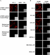
There is an increasing medical need to detect and spatially localize early and aggressive forms of prostate cancer. Affinity ligands derived from bacteriophage (phage) library screens can be developed to molecularly target prostate cancer with fluorochromes for optical imaging. Toward this goal, we used in vivo phage display and a newly described micropanning assay to select for phage that extravasate and bind human PC-3 prostate carcinoma xenografts in severe combined immune deficiency mice. One resulting phage clone (G1) displaying the peptide sequence IAGLATPGWSHWLAL was fluorescently labeled with the near-infrared fluorophore AlexaFluor 680 and was evaluated both in vitro and in vivo for its ability to bind and target PC-3 prostate carcinomas. The fluorescently labeled phage clone (G1) had a tumor-to-muscle ratio of approximately 30 in experiments. In addition, prostate tumors (PC-3) were readily detectable by optical-imaging methods. These results show proof of principle that disease-specific library-derived fluorescent probes can be rapidly developed for use in the early detection of cancers by optical means.
Figures


Similar articles
-
Streamlined in vivo selection and screening of human prostate carcinoma avid phage particles for development of peptide based in vivo tumor imaging agents.Comb Chem High Throughput Screen. 2011 Jan;14(1):9-21. doi: 10.2174/1386207311107010009. Comb Chem High Throughput Screen. 2011. PMID: 20958260
-
Bifunctional phage-based pretargeted imaging of human prostate carcinoma.Nucl Med Biol. 2009 Oct;36(7):789-800. doi: 10.1016/j.nucmedbio.2009.04.010. Epub 2009 Jul 9. Nucl Med Biol. 2009. PMID: 19720291 Free PMC article.
-
Novel α(2)β(1) integrin-targeted peptide probes for prostate cancer imaging.Mol Imaging. 2011 Aug;10(4):284-94. doi: 10.2310/7290.2010.00044. Epub 2011 Apr 1. Mol Imaging. 2011. PMID: 21486537
-
Phage Display's Prospects for Early Diagnosis of Prostate Cancer.Viruses. 2024 Feb 10;16(2):277. doi: 10.3390/v16020277. Viruses. 2024. PMID: 38400052 Free PMC article. Review.
-
Phage peptide display.Handb Exp Pharmacol. 2008;(185 Pt 2):145-63. doi: 10.1007/978-3-540-77496-9_7. Handb Exp Pharmacol. 2008. PMID: 18626602 Review.
Cited by
-
Optical tecnology developments in biomedicine: history, current and future.Transl Med UniSa. 2011 Oct 17;1:51-150. Print 2011 Sep. Transl Med UniSa. 2011. PMID: 23905030 Free PMC article.
-
Targeted nanoparticles for imaging incipient pancreatic ductal adenocarcinoma.PLoS Med. 2008 Apr 15;5(4):e85. doi: 10.1371/journal.pmed.0050085. PLoS Med. 2008. PMID: 18416599 Free PMC article.
-
Bacteriophage Capsid Modification by Genetic and Chemical Methods.Bioconjug Chem. 2021 Mar 17;32(3):466-481. doi: 10.1021/acs.bioconjchem.1c00018. Epub 2021 Mar 4. Bioconjug Chem. 2021. PMID: 33661607 Free PMC article. Review.
-
Discovery of novel peptides targeting pro-atherogenic endothelium in disturbed flow regions -Targeted siRNA delivery to pro-atherogenic endothelium in vivo.Sci Rep. 2016 May 12;6:25636. doi: 10.1038/srep25636. Sci Rep. 2016. PMID: 27173134 Free PMC article.
-
Ovarian Cancer Targeting Phage for In Vivo Near-Infrared Optical Imaging.Diagnostics (Basel). 2019 Nov 10;9(4):183. doi: 10.3390/diagnostics9040183. Diagnostics (Basel). 2019. PMID: 31717613 Free PMC article.
References
-
- Jemal A, Murray T, Ward E, Samuels A, Tiwari RC, Ghafoor A, Feuer EJ, Thun MJ. Cancer statistics, 2005. CA Cancer J Clin. 2005;55:10–30. - PubMed
-
- Centers for Disease Control, author. Fact sheet prostate cancer: The public health perspective. 2003 ( www.cdc.gov/cancer/prostate/prostate.htm)
-
- Poul MA, Becerril B, Nielsen UB, Morisson P, Marks JD. Selection of tumor-specific internalizing human antibodies from phage libraries. J Mol Biol. 2000;301:1149–1161. - PubMed
-
- Karasseva N, Glinsky VV, Chen NX, Komatireddy R, Quinn TP. Identification and characterization of peptides that bind human ErbB-2 selected from a bacteriophage display library. J Protein Chem. 2002;21:287–296. - PubMed
-
- Peletskaya EN, Glinsky VV, Glinsky GV, Deutscher SL, Quinn TP. Characterization of peptides that bind the tumor-associated Thomsen-Friedenreich antigen selected from bacteriophage display libraries. J Mol Biol. 1997;270:374–384. - PubMed
Publication types
MeSH terms
Grants and funding
LinkOut - more resources
Full Text Sources
Other Literature Sources
Medical
