. 2007 Jan 15;109(2):626-31.
doi: 10.1182/blood-2006-02-003665. Epub 2006 Sep 19.

Regulation of dendritic cell migration and adaptive immune response by leukotriene B4 receptors: a role for LTB4 in up-regulation of CCR7 expression and function

Affiliations

Regulation of dendritic cell migration and adaptive immune response by leukotriene B4 receptors: a role for LTB4 in up-regulation of CCR7 expression and function

Annalisa Del Prete et al. Blood. .

Abstract

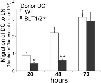
Trafficking of dendritic cells (DCs) to peripheral tissues and to secondary lymphoid organs depends on chemokines and lipid mediators. Here, we show that bone marrow-derived DCs (BM-DCs) express functional leukotriene B4 (LTB4) receptors as observed in dose-dependent chemotaxis and calcium mobilization responses. LTB4, at low concentrations, promoted the migration of immature and mature DCs to CCL19 and CCL21, which was associated with a rapid (30-minute) increase of CCR7 expression at the membrane level. At longer incubation times (6 hours), gene array analysis revealed a promoting role of LTB4, showing a significant increase of CCR7 and CCL19 mRNA levels. BM-DCs cultured from BLT1-/- or BLT1/2-/- mice showed a normal phenotype, but in vivo BLT1/2-/-DCs showed dramatic decrease in migration to the draining lymph nodes relative to wild-type (WT) DCs. Consistent with these observations, BLT1/2-/- mice showed a reduced response in a model of 2,4-dinitro-fluorobenzene (DNFB)-induced contact hypersensitivity. Adoptive transfer of 2,4-dinitrobenzene sulfonic acid (DNBS)-pulsed DCs directly implicated the defect in DC migration to lymph node with the defect in contact hypersensitivity. These results provide strong evidence for a role of LTB4 in regulating DC migration and the induction of adaptive immune responses.


Conflict of interest statement

Conflict-of-interest disclosure: The authors declare no competing financial interests.

Figures

Figure 1
Figure 1
Functional expression of LTB4 receptors on murine BM-DCs. Immature (iDCs) and mature DCs (20 ng/mL TNF-α for 24 hours) were generated from BM CD34+ precursors in vitro. Chemotaxis (left panel): Ligand-dependent chemotaxis of both iDCs and mDCs was measured using a 48-well micro chemotaxis chamber, as described in “Chemotaxis assay.” Data are the mean ± SE of cells from 3 individual fields for each concentration from a representative experiment of at least 3 repetitions. Calcium mobilization (right panel): Both iDCs and mDCs loaded with Indo-1 were induced with various concentrations of LTB4, and Ca2+ mobilization was measured. The y-axis indicates the fluorescence ratio (F340/380). Arrows indicate LTB4 addition.
Figure 2
Figure 2
Characterization of BM-derived WT and BLT1/2−/− DCs. (A) FACS analysis of membrane phenotype in WT and BLT1/2−/− DCs. Gray line indicates isotype control; broken line, iDCs; and black line, mDCs. (B) FITC-dextran uptake. (C) Mixed leukocyte reaction induced by WT and BLT1/2−/− DCs. (D,E) Calcium mobilization measurement in response to LTB4 in WT (D) and in BLT1/2−/− (E) DCs. (F, G) In vitro chemotaxis of iDCs (F) and mDCs (G) toward CCL3 (100 ng/mL), CCL19 (100 ng/mL), and LTB4 (100 nM). Error bars indicate SE.
Figure 3
Figure 3
Chemotaxis of LTB4-pretreated DCs. BM-derived WT DCs, both iDCs (A) and mDCs (B), were pretreated with LTB4 as indicated for 30 minutes at 37°C. The chemotactic response toward a fixed concentration (100 ng/mL) of CCL3 and CCL19 (iDCs) and toward CCL19 and CCL21 (mDCs) was determined. Significant difference in chemotaxis levels between untreated and LTB4-treated DCs was indicated (*P < .05, *P < .01 by t test). Data are the mean ± SE of cells from 3 individual fields for each concentration from a representative experiment of at least 3 repetitions. (C) Checkerboard analysis of DC migration across polycarbonate filters toward CCL19.
Figure 4
Figure 4
Effect of LTB4 treatment on surface expression of CCR7. (A) FACS analysis of CCR7 expression on the surface and after permeabilization in iDCs after 30 minutes of LTB4 (1 nM) treatment. Gray line indicates isotype control; broken line, iDCs; and black line, LTB4-treated iDCs. (B) CCL19-Fc fusion protein staining of untreated and LTB4-treated iDCs, both before and after permeabilization, analyzed by confocal microscopy (×100).
Figure 5
Figure 5
LTB4-induced changes in gene expression profiles. A cytokine and chemokine SuperArray was performed using 2 μg/mL RNA for each sample as described in “Methods.” The graph indicates the fold increase of each group over iDCs showing changes in CCR1, CCR7, and CCL19.
Figure 6
Figure 6
Defective in vivo migration of BM-BLT1/2−/− DCs. Mature DCs from WT and BLT1/2−/− mice were labeled with the vital dye CFSE and injected subcutaneously in the hind leg footpad of WT mice. Popliteal lymph nodes were recovered at the indicated times and the cell suspension was evaluated by flow cytometry. One experiment representative of 3 is shown (*P < .05, *P < .01 by t test, vs respective WT control group). Error bars indicate SE.
Figure 7
Figure 7
Defective CHS in BLT1/2−/− mice. (A) WT and BLT1−/− mice were sensitized on the shaved abdominal skin with 50 μL of 0.5% DNFB in 4:1 acetone/olive oil (vol/vol) and 5 μL on each footpad. Five days later, mice were challenged on the right ear (10 μL of 0.2% DNFB on each side). The left ear was painted with vehicle as control. Increases in ear swelling were measured at different time points. Mean values ± SE for each group (5 mice) are presented. (*P < .05, **P < .01 by t test, vs respective WT control group). One experiment representative of 3 is shown. (B) Histology, hematoxylin and eosin–stained tissue sections from WT and BLT1/2−/− mice after 24 hours read-out CHS. The top row represents a ×100 magnification; an enlarged region of the same pictures is shown in the bottom row. Images were acquired using a Carl Zeiss Axioskope Optical Microscope equipped with an Achroplan 10 ×/0.25 NA objective and a 10 × eyepiece. Images were captured using a Nikon CoolPix 3.34 Megapixel camera (Nikon, Tokyo, Japan) at a fine resolution of 1600 × 1200 pixels in JPEG format and processed using Adobe Photoshop (Adobe Systems, San Jose, CA). (C) Defective capacity of BLT1/2−/− DCs to elicit CHS. WT mice were immunized (on day −5) by subcutaneous injection of DNBS-loaded DCs (106/mouse) obtained from WT or BLT1/2−/− mice. Mice were challenged 5 days later (day 0) by ear painting with DNFB. Figure represents the values for ear swelling (mean ± SE) of 5 mice per group, and is representative of 3 experiments. **P < .01 by t test.

References

    1. Banchereau J, Briere F, Caux C, et al. Immunobiology of dendritic cells. Annu Rev Immunol. 2000;18:767–811. - PubMed
    1. Steinman RM, Hawiger D, Nussenzweig MC. Tolerogenic dendritic cells. Annu Rev Immunol. 2003;21:685–711. - PubMed
    1. Dieu MC, Vanbervliet B, Vicari A, et al. Selective recruitment of immature and mature dendritic cells by distinct chemokines expressed in different anatomic sites. J Exp Med. 1998;188:373–386. - PMC - PubMed
    1. Sozzani S, Allavena P, D'Amico G, et al. Differential regulation of chemokine receptors during dendritic cell maturation: a model for their trafficking properties. J Immunol. 1998;161:1083–1086. - PubMed
    1. MartIn-Fontecha A, Sebastiani S, Hopken UE, et al. Regulation of dendritic cell migration to the draining lymph node: impact on T lymphocyte traffic and priming. J Exp Med. 2003;198:615–621. - PMC - PubMed

Publication types