

Case Reports
Spontaneous retroperitoneal hemorrhage caused by segmental arterial mediolysis
- PMID: 16985559
- PMCID: PMC1471764
Item in Clipboard
Case Reports
Spontaneous retroperitoneal hemorrhage caused by segmental arterial mediolysis
Rev Urol.
2006 Winter.
Abstract
Spontaneous retroperitoneal hemorrhage is a rare clinical entity; signs and symptoms include pain, hematuria, and shock. Spontaneous retroperitoneal hemorrhage can be caused by tumors, such as renal cell carcinoma and angiomyolipoma; polyarteritis nodosa; and nephritis. The least common cause is segmental arterial mediolysis. Although computed tomography is used for the diagnosis of spontaneous retroperitoneal hemorrhage, it can miss segmental arterial mediolysis as the cause of the hemorrhage. The diagnosis of segmental arterial mediolysis as a cause of spontaneous retroperitoneal hemorrhage requires angiography, with pathologic confirmation for a definitive diagnosis.
Figures
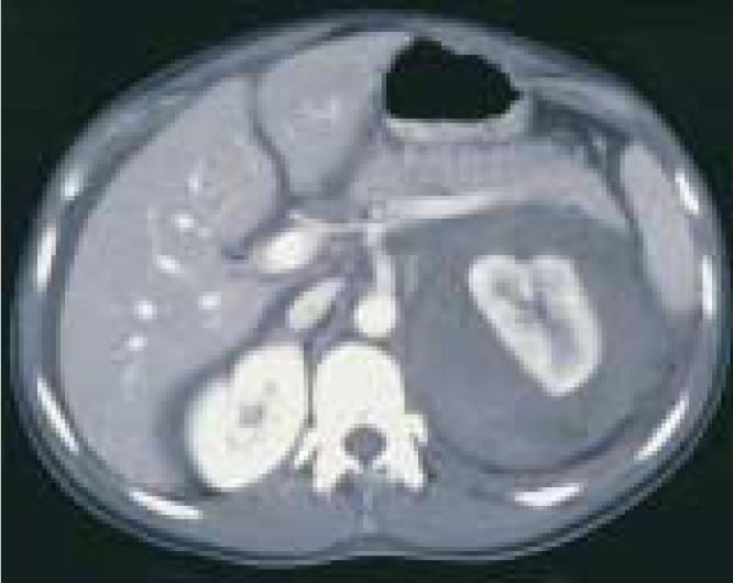
CT scan, obtained the day after admission because the patient’s abdomen became distended, revealed a large left perinephric hematoma.
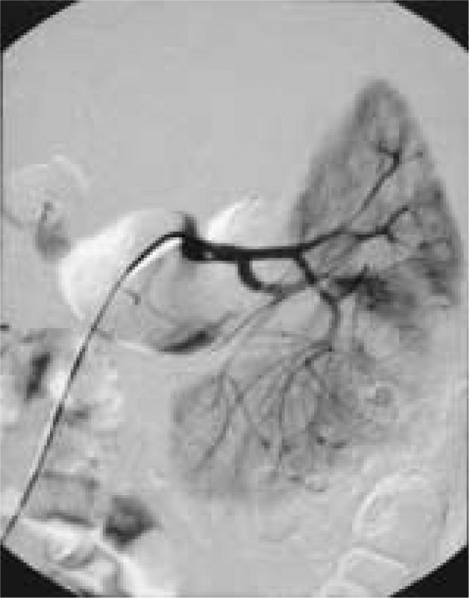
Initial angiography failed to demonstrate any active bleeding, but several microaneurysms were seen in the peripheral regions of the renal cortex. These aneurysms are usually suggestive of polyarteritis nodosa.
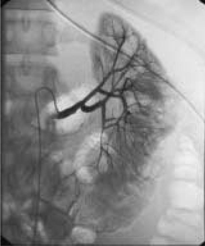
Repeat angiography performed 2 days after the initial on showed that the patients’s microaneurysms had been replaced by several blind-ending vessels. The patient’s right kidney and celiac axis were normal (not shown).
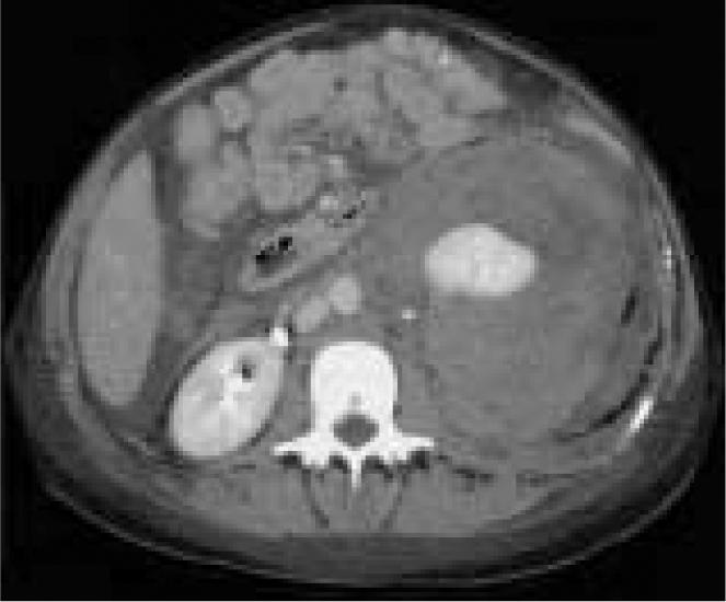
Repeat CT scan obtained 4 days after the patient’s initial CT showed that the perinephric hematoma had increased from 6 to 10.25 cm and extended into the pelvis. Additional cuts revealed free blood in the peritoneum and a infarct of the left kidney.
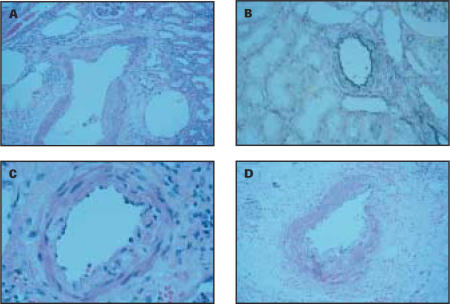
Pathology of the patient’s kidney revealed segmental arterial mediolysis, which is characterized by patch, non-circumferential loss of the media (A) and the external elastic lamina (B). Weakening of the media is exacerbated by focal vacuolization of this layer (C). Inflammation has been described in this condition sporadically and was seen in this patient (D).
References
-
- Polkey HJ, Vynalek WJ. Spontaneous nontraumatic perirenal and renal hematomas. Arch Surg. 1933;26:196.
-
- Wolff JM, Jung PK, Adam G, Jakse G. Spontaneous retroperitoneal haemorrhage associated with renal disease. J R Coll Surg Edinb. 1998;43:53–56. - PubMed
-
- McDougal WS, Kursh ED, Persky L. Spontaneous rupture of the kidney with perirenal hematoma. J Urol. 1975;114:181–184. - PubMed
-
- Morgentaler A, Belville JS, Tumeh SS, et al. Rational approach to evaluation and management of spontaneous perirenal hemorrhage. Surg Gynecol Obstet. 1990;170:121–125. - PubMed
-
- Chang S, Ma C, Lee S. Spontaneous retroperitoneal hemorrhage from kidney causes. Eur Urol. 1988;15:281–284. - PubMed
Publication types
LinkOut - more resources
Full Text Sources