

Prostatic intraepithelial neoplasia: an overview
Abstract
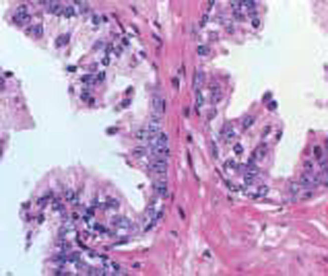
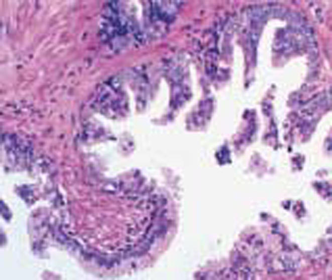
Prostatic intraepithelial neoplasia (PIN) is the most established precursor of prostatic carcinoma. The presence of prominent nucleoli within an existing duct structure is an easy way to identify the disorder. Four main patterns of high-grade PIN (HGPIN) have been described: tufting, micropapillary, cribriform, and flat. In addition to exhibiting similar cytologic features, both HGPIN and prostatic carcinoma are associated with increased incidence and severity with age, and with high rates of occurrence in the peripheral zone of the prostate. HGPIN and prostate cancer share genetic and molecular markers as well, with PIN representing an intermediate stage between benign epithelium and invasive malignant carcinoma. The clinical significance of HGPIN is that it identifies patients at risk for malignancy. With the increased use of extended biopsy protocols, clinicians are more likely to identify HGPIN and less likely to miss concurrent carcinoma. Androgen deprivation therapy decreases the prevalence and extent of PIN, and may play a role in chemoprevention. Preliminary studies suggest that selective estrogen receptor modulators may also prevent the progression of HGPIN to prostate cancer.
Figures


Similar articles
-
Prostatic intraepithelial neoplasia: recent advances.Arch Pathol Lab Med. 2007 Aug;131(8):1257-66. doi: 10.5858/2007-131-1257-PINRA. Arch Pathol Lab Med. 2007. PMID: 17683188 Review.
-
[Diagnostic criteria and differential diagnosis of prostatic intraepithelial neoplasia].Pathologe. 1998 Jan;19(1):33-41. doi: 10.1007/s002920050252. Pathologe. 1998. PMID: 9541940 Review. German.
-
High-grade prostatic intraepithelial neoplasia on needle biopsy: risk of cancer on repeat biopsy related to number of involved cores and morphologic pattern.Am J Surg Pathol. 2004 May;28(5):629-33. doi: 10.1097/00000478-200405000-00010. Am J Surg Pathol. 2004. PMID: 15105651
-
High-grade prostatic intraepithelial neoplasia, PIN-like carcinoma, ductal carcinoma, and intraductal carcinoma of the prostate.Mod Pathol. 2018 Jan;31(S1):S71-79. doi: 10.1038/modpathol.2017.138. Mod Pathol. 2018. PMID: 29297491 Review.
-
Histological markers of risk and the role of high-grade prostatic intraepithelial neoplasia.Urology. 2001 Apr;57(4 Suppl 1):115-20. doi: 10.1016/s0090-4295(00)00953-5. Urology. 2001. PMID: 11295607 Review.
Cited by
-
Pericyte in Oral Squamous Cell Carcinoma: A Systematic Review.Head Neck Pathol. 2020 Dec;14(4):1080-1091. doi: 10.1007/s12105-020-01188-2. Epub 2020 Jun 6. Head Neck Pathol. 2020. PMID: 32506378 Free PMC article.
-
FRMD6 has tumor suppressor functions in prostate cancer.Oncogene. 2021 Jan;40(4):763-776. doi: 10.1038/s41388-020-01548-w. Epub 2020 Nov 28. Oncogene. 2021. PMID: 33249427
-
Reduced cytoplasmic expression of MAGE-A2 predicts tumor aggressiveness and survival: an immunohistochemical analysis.World J Urol. 2021 Jun;39(6):1831-1843. doi: 10.1007/s00345-020-03395-6. Epub 2020 Aug 9. World J Urol. 2021. PMID: 32772147
-
The helix-loop-helix transcriptional regulator Id4 is required for terminal differentiation of luminal epithelial cells in the prostate.Oncoscience. 2021 Mar 24;8:14-30. doi: 10.18632/oncoscience.524. eCollection 2021. Oncoscience. 2021. PMID: 33884281 Free PMC article.
-
MicroRNA-27a-5p regulation by promoter methylation and MYC signaling in prostate carcinogenesis.Cell Death Dis. 2018 Feb 7;9(2):167. doi: 10.1038/s41419-017-0241-y. Cell Death Dis. 2018. PMID: 29415999 Free PMC article.
References
-
- Jemal A, Tiwari RC, Murray T, et al. Cancer statistics. 2004. CA Cancer J Clin. 2004;54:8–29. - PubMed
-
- Bostwick DG, Brawer MK. Prostatic intraepithelial neoplasia and early invasion in prostate cancer. Cancer. 1987;59:788–794. - PubMed
-
- Kim HL, Yang XJ. Prevalence of high-grade prostatic intraepithelial neoplasia and its relationship to serum prostate specific antigen. Int Braz J Urol. 2002;28:413–417. - PubMed
-
- McNeal JE, Bostwick DG. Intrauctal dysplasia: a premalignant lesion of the prostate. Hum Pathol. 1986;17:64–71. - PubMed
-
- Epstein JL, Carmichael M, Partin AW. OA-519 (fatty acid synthase) as an independent predictor of pathologic state in adenocarcinoma of the prostate. Urology. 1995;45:81–86. - PubMed
LinkOut - more resources
Full Text Sources