. 2002;4 Suppl 4(Suppl 4):S7-S18.

Pathophysiology of overactive bladder and urge urinary incontinence

Pathophysiology of overactive bladder and urge urinary incontinence

William D Steers. Rev Urol. 2002.

Abstract

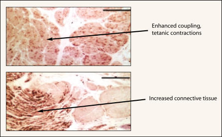
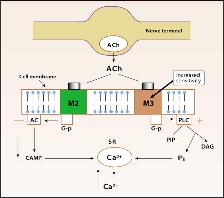
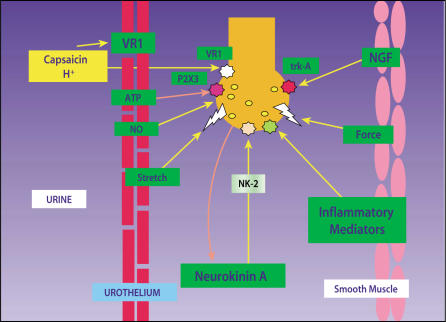
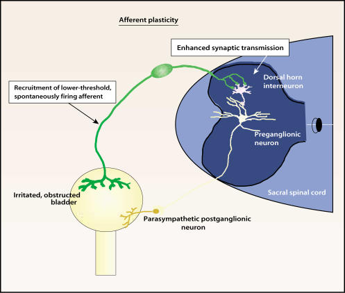
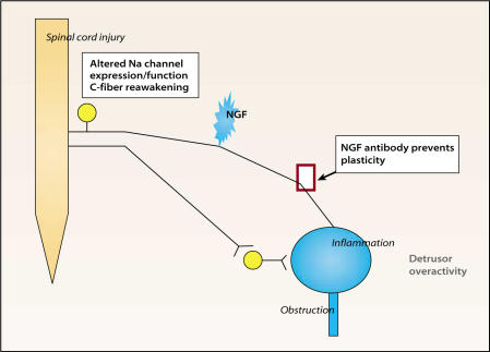
Storage symptoms such as urgency, frequency, and nocturia, with or without urge incontinence, are characterized as overactive bladder (OAB). OAB can lead to urge incontinence. Disturbances in nerves, smooth muscle, and urothelium can cause this condition. In some respects the division between peripheral and central causes of OAB is artificial, but it remains a useful paradigm for appreciating the interactions between different tissues. Models have been developed to mimic the OAB associated with bladder instability, lower urinary tract obstruction, neuropathic disorders, diabetes, and interstitial cystitis. These models share the common features of increased connectivity and excitability of both detrusor smooth muscle and nerves. Increased excitability and connectivity of nerves involved in micturition rely on growth factors that orchestrate neural plasticity. Neurotransmitters, prostaglandins, and growth factors, such as nerve growth factor, provide mechanisms for bidirectional communication between muscle or urothelium and nerve, leading to OAB with or without urge incontinence.


Figures

Figure 1
Figure 1
Bladder smooth muscle from patients in unstable bladder demonstrates increased myogenic activity, with fused tetanic contractions and changes in structure. Increased connective tissue between muscle fascicles is reminiscent of that seen with aging, obstruction, and apoptosis due to ischemia. Thus, myogenic changes are commonly seen in the OAB with or without urge incontinence. Reprinted from Morrison J, Steers WD, Brading A, et al. Neurophysiology and neuropharmacology. In: Khoury S, Abrams P, Wein A (eds). Incontinence, 2nd ed. Plymouth, UK: Health Publication Ltd; 2002.
Figure 2
Figure 2
Altered responses to stimuli occur in unstable bladders. Obstructed bladders demonstrate hypersensitivity to cholinergic agonists acting at muscarinic (M2 or M3) receptors and increased contractions due to potassium chloride, but reduced electrical evoked contractions. Detrusor tissue from idiopathic instability patients shows increased electrical evoked contractions but normal sensitivity to muscarinic agonists (M3 and M2). Denervated bladders show increased M3 receptor expression. Acetylcholine released from parasympathetic nerves supplying the bladder cause activation of M3 receptors responsible for bladder contraction in humans. The M3 elicited contraction is due to a rise in cytosolic calcium (Ca+2) from intracellular stores. Ca+2 is released from these stores following M3-coupled activation of G-protein (G-p) mediated phospholipase C (PLC) breakdown. Inositol triphosphate (IP3) triggers Ca+2 release from sacroplasmic reticulum (SR). M2 activation causes a fall in cyclic adenosine monophosphate (cAMP), preventing relaxation. Newer anticholinergics selectively target M3 receptors in the hope of reducing side effects and perhaps increasing efficacy. Reprinted from Morrison J, Steers WD, Brading A, et al. Neurophysiology and neuropharmacology. In: Khoury S, Abrams P, Wein A (eds). Incontinence, 2nd ed. Plymouth, UK: Health Publication Ltd; 2002.
Figure 3
Figure 3
The urothelium directly communicates with suburothelial afferents acting as luminal sensors. Increased sensitivity of these afferents may lead to overactive bladder (OAB). Low pH, high potassium concentration in the urine, and increased osmolality can influence sensory nerves through mediators such as nitric oxide (NO) and neurokinin A, which acts through NK-2 receptors. H+ ions activate vanilloid (VR-1) receptors, causing pain. Adenosine triphosphate (ATP) can activate purinergic P2X3 receptors and modulate sensation. Mice lacking the P2X3 receptor exhibit enlarged bladders and reduced sensation. Inflammatory cytokines and nerve growth factor (NGF) acting at tyrosine kinase A (trk A) receptors can influence the growth, transmitter synthesis, and function of neurons. Sensitization of suburothelial afferents without changes in smooth muscle may result only in urgency. If afferents are sensitized (lower thresholds, spontaneous burst firing) and smooth muscle coupling is enhanced, urgency with unstable contractions may result. Vanilloid, neurokinin, and purinergic antagonists are being examined as possible therapies for OAB and urge incontinence. Reprinted from Morrison J, Steers WD, Brading A, et al. Neurophysiology and neuropharmacology. In: Khoury S, Abrams P, Wein A (eds). Incontinence, 2nd ed. Plymouth, UK: Health Publication Ltd; 2002.
Figure 4
Figure 4
Sensitization of bladder afferents such as occurs following obstruction or inflammation may lead to enhanced transmission involving second-order neurons in the sacral spinal cord. If preganglionic neurons are not activated, then urgency and frequency without unstable contractions may result. Reprinted from Morrison J, Steers WD, Brading A, et al. Neurophysiology and neuropharmacology. In: Khoury S, Abrams P, Wein A (eds). Incontinence, 2nd ed. Plymouth, UK: Health Publication Ltd; 2002.
Figure 5
Figure 5
After transient environmental (ie, intravesical) changes such as inflammation or temporary obstruction, afferents may revert to normal activity. However, if patient is genomically predisposed to OAB (eg, exhibits familial urge incontinence or chronic pain syndromes such as interstitial cystitis, irritable bowel syndrome, or fibromyalgia) or long-term environmental changes occur, nerve growth factor (NGF) may alter afferents irreversibly. Antibody to NGF or fusion protein against the NGF receptor prevents overactive bladder (OAB) in animal models coincident with alterations in afferents. NGF’s long-term actions may rely on changes in sodium (Na+) channel isoforms expressed by afferents that influence excitability. Most unmyelinated (C-fiber) afferents are not responsive to normal stimuli such as distention or presence of intravesical contents. However, following spinal cord injury, obstruction, or inflammation, activation of the silent C-fibers occurs. The reawakening of silent C-fibers may correspond to changes in Na+ channel expression. Novel approaches to OAB in the future may target the mechanisms leading to these long-term changes. Reprinted from Morrison J, Steers WD, Brading A, et al. Neurophysiology and neuropharmacology. In: Khoury S, Abrams P, Wein A (eds). Incontinence, 2nd ed. Plymouth, UK: Health Publication Ltd; 2002.

References

    1. Eckhardt MD, van Venrooij GE, Boon TA. Symptoms, prostate volume, and urodynamic findings in elderly male volunteers without and with LUTS and in patients with LUTS suggestive of benign prostatic hyperplasia. Urology. 2001;58:966–971. - PubMed
    1. Sibley GNA. The Response of the Bladder to Lower Urinary Tract Obstruction [DM thesis] Oxford, England: Oxford University; 1984.
    1. German K, Bedwani J, Davies J, et al. What is the pathophysiology of detrusor hyperreflexia? Neurourol Urodyn. 1993;12:335–336.
    1. Sibley GNA. Developments in our understanding of detrusor instability. Br J Urol. 1997;80:54–61. - PubMed
    1. Brading AF, Turner WH. The unstable bladder: towards a common mechanism. Br J Urol. 1994;73:3–8. - PubMed

LinkOut - more resources