. 2007 Jan 1;401(1):205-16.
doi: 10.1042/BJ20061128.

Yeast sphingolipids do not need to contain very long chain fatty acids

Affiliations

Yeast sphingolipids do not need to contain very long chain fatty acids

Vanessa Cerantola et al. Biochem J. .

Abstract

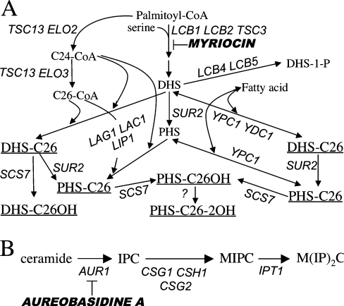
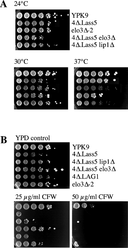
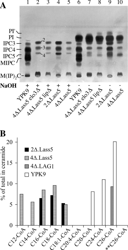
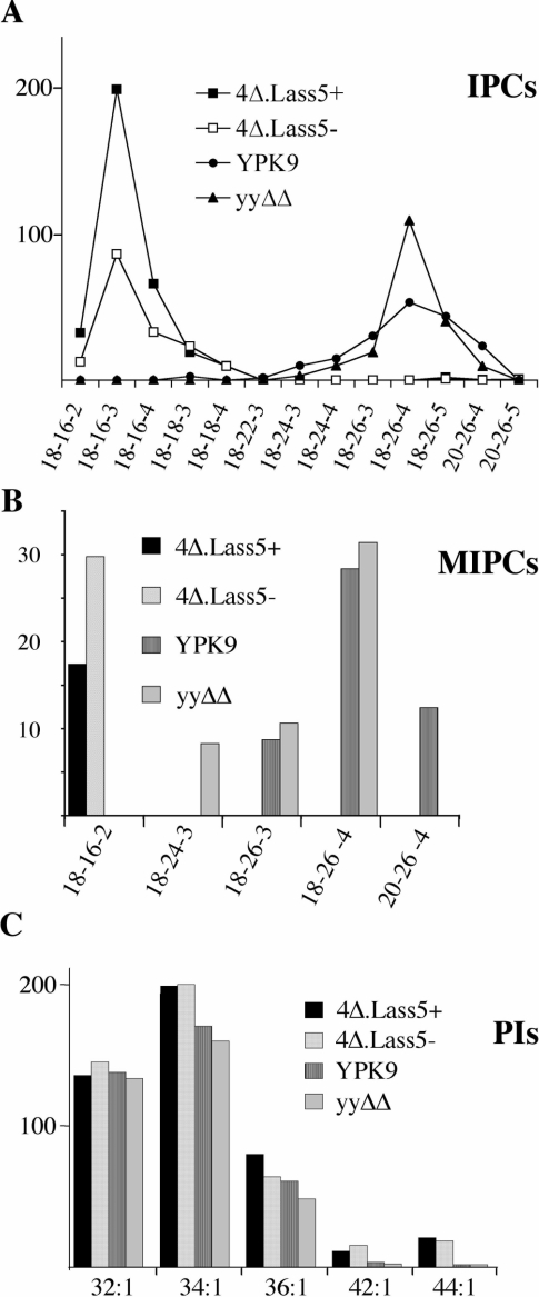
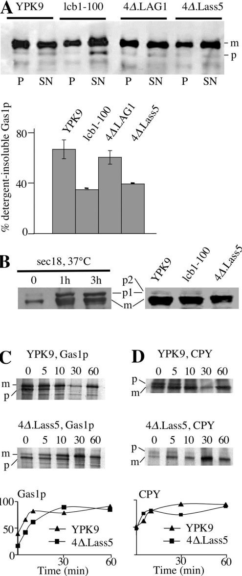
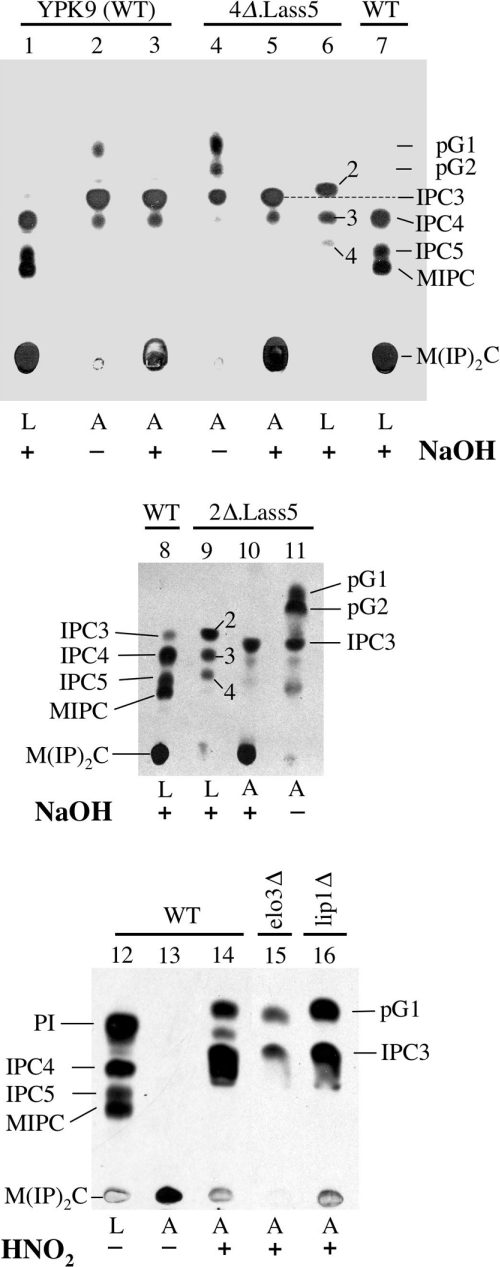
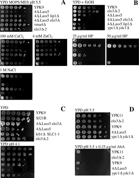
Synthesis of VLCFAs (very long chain fatty acids) and biosynthesis of DHS (dihydrosphingosine) both are of vital importance for Saccharomyces cerevisiae. The bulk of VLCFAs and DHS are used for ceramide synthesis by the Lag1p (longevity-assurance gene 1)/Lac1p (longevity-assurance gene cognate 1)/Lip1p (Lag1p/Lac1p interacting protein) ceramide synthase. LAG1 and LAC1 are redundant but LIP1 is essential. Here we show that 4Delta (lag1Deltalac1Deltaypc1Deltaydc1Delta) cells devoid of all known endogenous ceramide synthesis pathways are unviable but can be rescued by the expression of Lass5, a mouse LAG1 homologue. Ceramide synthase activity of 4Delta.Lass5 cells only utilizes C16 and C18 fatty acids and does not require the help of Lip1p, an essential cofactor of Lag1p/Lac1p. HPLC-electrospray ionization-MS/MS analysis demonstrated that in IPCs (inositolphosphorylceramides) of 4Delta.Lass5, the very long chain fatty acids (C26 and C24) account for <1% instead of the normal >97%. Notwithstanding, IPCs incorporated into glycosylphosphatidylinositol anchors of 4Delta.Lass5 show normal mobility on TLC and the ceramide- and raft-dependent traffic of Gas1p (glycophospholipid-anchored surface protein) from endoplasmic reticulum to Golgi remains almost normal. Moreover, the biosynthesis of C24:0 fatty acids remains essential. Thus, C(24:0) and dihydrosphingosine are both necessary for survival of yeast cells even if they utilize C16 and C18 fatty acids for sphingolipid biosynthesis.


Figures

Figure 1
Figure 1. Sphingolipid biosynthesis in Saccharomyces cerevisiae
(A) Biosynthesis of ceramides (underlined). (B) Generation of mature sphingolipids from ceramide. Gene names and inhibitors are in italics.
Figure 2
Figure 2. Temperature and Calcofluor White sensitivity of 4Δ.Lass5
Serial 10-fold dilutions of cells were plated on YPD + uracil + adenine agar plates. (A) Plates were incubated at 24, 30 or 37 °C. (B) Plates contained 0, 25 or 50 μg/ml of Calcofluor White (CFW) and were incubated at 30 °C. All plates were photographed after 3 days.
Figure 3
Figure 3. 4Δ.Lass5 make abnormal IPCs and use C16– and C18–CoA for ceramide synthesis
(A) The indicated strains were labelled with [3H]inositol, washed, and lipids (plus intracellular free [3H]inositol) were extracted. Aliquots of extract corresponding to 106 c.p.m. (counts per min) were treated with NaOH (lanes 1–5) or control incubated (lanes 6–10) and desalted lipids were separated by TLC in solvent system 2. IPC2, IPC3, IPC4 and IPC5 and numbers 2, 3 and 4 next to lane 2 designate types of IPC, which contain a total number of 2 to 5 hydroxy groups in their ceramide moiety. The numbers next to lane 2 are rendered likely by HPLC–ESI-MS/MS results (Figure 4 and supplementary Figure S1). (B) Microsomes of 4Δ.Lass5 were radiolabelled for 2 h with [3H]DHS in the presence of the indicated acyl-CoAs (0.1 mM) or free fatty acids (0.1 mM) for control. The radiolabelled lipids were extracted, analysed by TLC using solvent system 1 and quantitated by radioscanning. Counts present in ceramide were given as the percentage of total counts present in the lane. No ceramides were made when microsomes were incubated with free fatty acids (results not shown). ** Tubes containing boiled microsomes were used as a further control.
Figure 4
Figure 4. Sphingolipids from 4Δ.Lass5 cells contain mainly C16:0 and C18:0 fatty acids by HPLC–ESI-MS/MS
All sphingolipid classes were extracted quantitatively [35] from cells growing exponentially in LM at 30 °C and analysed by HPLC–ESI-MS/MS. YPK9, ypc1Δ ydc1Δ (yyΔΔ) and 4Δ.Lass5 cells were all grown with methionine (4Δ.Lass5+), 4Δ.Lass5 also without methionine (4Δ.Lass5−). Parent ion and fragment ion masses (m/z) were recorded throughout the HPLC run and were screened for all theoretically possible sphingolipids containing different saturated, monounsaturated, mono- or di-hydroxylated fatty acids combined with DHS or PHS as well as for conventional phospholipids [40]. The vertical axis in all panels gives the ion counts in thousands. All species shown in (A) were identified as phosphoinositides by characteristic fragment ions at m/z 259 and 241, corresponding to inositolphosphate and inositolphosphate–H2O, except for those that were less than 1% of the most predominant IPC. However, the LCB could not be identified by fragmentation and sphingolipids were annotated by three consecutive numbers to indicate the probable number of carbon atoms in the LCB, the probable number of carbon atoms in the fatty acid, and the sum of hydroxy groups in the ceramide moiety respectively. (A) The lipid extracts were mixed with lipid extracts from WT cells grown in [13C]glucose as the only carbon source and the results were normalized using this internal standard. The normalized number of ions derived from IPCs present in extracts of 4Δ.Lass5+, 4Δ.Lass5− and ypc1Δ ydc1Δ amounted to 183%, 92% and 106% of the WT (YPK9) respectively. (C) The PIs are specified by two consecutive numbers indicating the total number of carbon atoms and double bonds present in the two fatty acids respectively. The Figure shows one representative of several experiments yielding very similar results.
Figure 5
Figure 5. Raft association and transport of Gas1p in 4Δ.Lass5
(A) 4Δ.Lass5 was grown in LM (His−, Met−), 4Δ.LAG1 in LM (−ura) containing galactose instead of glucose, YPK9 and lcb1-100 in YPD. All cells were grown at 30 °C except for lcb1-100, which was kept at 24 °C. Exponentially growing cells were lysed with glass beads and aliquots of lysate were solubilized in Triton X-100 for a membrane association assay (see the Materials and methods section). Triton X-100 lysates were subjected to ultracentrifugation, the soluble (SN) and the pellet (P) fractions were processed for Western blotting using anti-Gas1p (A) or anti-CPY antibodies (B), allowing the detection of mature (m) and precursor (p) forms. (A) Quantitation of results of top panel and of a second identical experiment is also shown. In (B) sec18Δ cells were incubated at 37 °C for 0, 1 and 3 h before lysis. For kinetic analysis of Gas1p (C) and CPY (D) maturation, yeast cells were metabolically labelled with [35S]methionine and [35S]cysteine, the label was chased for 0–60 min and Gas1p and CPY were immunoprecipitated from cell extracts, resolved by SDS/PAGE, and detected by phosphoimaging and by fluorography. Relative amounts of mature proteins are indicated in bottom panels.
Figure 6
Figure 6. GPI anchor lipids of WT and mutant 4Δ.Lass5 cells are similar
Labelling of 25 A600 units of YPK9 (WT), 4Δ.Lass5, 2Δ.Lass5, 4Δ.Lass5 elo3Δ (lane 15) and 4Δ.Lass5 lip1Δ (lane 16) cells for 2 h with 100 μCi of [3H]inositol. Free lipids were extracted, GPI proteins were completely delipidated and their lipid moieties were liberated using HNO2, with the exception of lane 13. Free lipids (L, lanes 1, 6–9 and 12) and liberated anchor lipids (A, lanes 2–5, 10, 11 and 13–16) were treated with NaOH (+) or mock incubated (−) for deacylation, separated by TLC and exposed for fluorography. IPCs were annotated as in Figure 3(A).
Figure 7
Figure 7. Phenotypic analysis of 4Δ.Lass5 cells under various stress conditions
Serial 10-fold dilutions of cells were plated on buffered (A and D) or unbuffered YPD + uracil + adenine plates containing the indicated ingredients. Plates were then incubated at 30 °C and photographed after 3 days except for panel (D), where they were grown at 24 °C for 3 days. 4Δ.LAG1 cells were grown in YPG before being plated on YPD, where they strongly reduce sphingolipid biosynthesis but, nevertheless, continue to grow for several days. The vma4Δ cells served as a positive control and exhibited the same Vma phenotype as described for vma2Δ and vma13Δ [23].

References

    1. Dickson R. C., Sumanasekera C., Lester R. L. Functions and metabolism of sphingolipids in Saccharomyces cerevisiae. Prog. Lipid Res. 2006;45:447–465. - PubMed
    1. Smith S. W., Lester R. L. Inositol phosphorylceramide, a novel substance and the chief member of a major group of yeast sphingolipids containing a single inositol phosphate. J. Biol. Chem. 1974;249:3395–3405. - PubMed
    1. Oh C. S., Toke D. A., Mandala S., Martin C. E. ELO2 and ELO3, homologues of the Saccharomyces cerevisiae ELO1 gene, function in fatty acid elongation and are required for sphingolipid formation. J. Biol. Chem. 1997;272:17376–17384. - PubMed
    1. Sims K. J., Spassieva S. D., Voit E. O., Obeid L. M. Yeast sphingolipid metabolism: clues and connections. Biochem. Cell Biol. 2004;82:45–61. - PubMed
    1. Horvath A., Sutterlin C., Manning-Krieg U., Movva N. R., Riezman H. Ceramide synthesis enhances transport of GPI-anchored proteins to the Golgi apparatus in yeast. EMBO J. 1994;13:3687–3695. - PMC - PubMed

Publication types