Review
. 2006 Oct 15;399(2):177-90.
doi: 10.1042/BJ20060854.

Structure, function and mechanism of exocyclic DNA methyltransferases

Affiliations
Review

Structure, function and mechanism of exocyclic DNA methyltransferases

Shivakumara Bheemanaik et al. Biochem J. .

Erratum in

  • Biochem J. 2006 Nov 1;399(3):543

Abstract

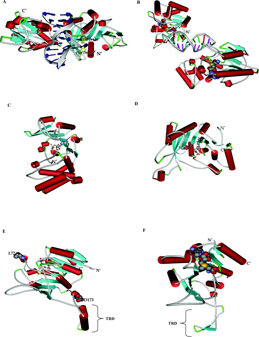
DNA MTases (methyltransferases) catalyse the transfer of methyl groups to DNA from AdoMet (S-adenosyl-L-methionine) producing AdoHcy (S-adenosyl-L-homocysteine) and methylated DNA. The C5 and N4 positions of cytosine and N6 position of adenine are the target sites for methylation. All three methylation patterns are found in prokaryotes, whereas cytosine at the C5 position is the only methylation reaction that is known to occur in eukaryotes. In general, MTases are two-domain proteins comprising one large and one small domain with the DNA-binding cleft located at the domain interface. The striking feature of all the structurally characterized DNA MTases is that they share a common core structure referred to as an 'AdoMet-dependent MTase fold'. DNA methylation has been reported to be essential for bacterial virulence, and it has been suggested that DNA adenine MTases (Dams) could be potential targets for both vaccines and antimicrobials. Drugs that block Dam could slow down bacterial growth and therefore drug-design initiatives could result in a whole new generation of antibiotics. The transfer of larger chemical entities in a MTase-catalysed reaction has been reported and this represents an interesting challenge for bio-organic chemists. In general, amino MTases could therefore be used as delivery systems for fluorescent or other reporter groups on to DNA. This is one of the potential applications of DNA MTases towards developing non-radioactive DNA probes and these could have interesting applications in molecular biology. Being nucleotide-sequence-specific, DNA MTases provide excellent model systems for studies on protein-DNA interactions. The focus of this review is on the chemistry, enzymology and structural aspects of exocyclic amino MTases.


Figures

Figure 1
Figure 1. Arrangement of conserved motifs in the primary structure of exocyclic DNA MTases
Figure 2
Figure 2. Comparison of the topologies and location of conserved motifs in DNA MTases of different families
Locations of the conserved motifs displayed on the framework of the structure of the large domain of C5-cytosine MTases. Topology and locations of important amino acid residues in different MTases are shown [5]: C5-cytosine MTases (A), α-class MTases (B), β-class MTases (C), and γ-class MTases (D). Adapted from [5] by permission of Wiley-VCH and the author.
Figure 3
Figure 3. Proposed reaction mechanisms for exocyclic DNA methylation
(A) Amino methylation via cationic intermediate [17]. (B) Proposed reaction mechanism for N4-cytosine MTases such as M.PvuII.
Figure 4
Figure 4. Sequential kinetic mechanisms observed with AdoMet-dependent DNA MTases
(A) Random Bi Bi mechanism. (B) Ordered mechanism, AdoMet binding first. (C) Ordered mechanism, DNA binding first. meDNA, methylated DNA.
Figure 5
Figure 5. Structures of exocyclic DNA MTases
(A) Three-dimensional structure of M.TaqI with DNA (PDB code 1G38). The DNA backbone in the structure is shown as arrows and base pairs are shown as blue rings. The nucleotide shown in blue and present within the protein back bone is the flipped target adenine. Tyr108 and Phe196 shown in yellow are present in the vicinity of flipped target adenine. (B) The crystal structure of two molecules of T4 Dam bound to a palindromic 12 bp DNA duplex in presence of AdoHcy (PDB code 1Q0T). The DNA backbone in the structure is shown as tubes, base pairs are shown as sticks and AdoHcy molecules are shown as space-filled models. (C) The crystal structure of DpnM with AdoMet (PDB code 2DPM). The AdoMet molecule in the structure is shown as a ball and stick model. (D) Three-dimensional structure of M.PvuII in presence of AdoHcy (PDB code 1B00). The AdoHcy molecule in the structure is shown as a ball and stick representation. (E) The crystal structure of M.RsrI in presence of AdoMet (PDB code 1NW5). The AdoMet molecule in the structure is shown as a ball and stick model. The two amino acids (Leu72 and Asp173) are represented as space-fill models. (F) The crystal structure of MboIIA in the presence of AdoMet (PDB code 1G60). The AdoMet molecule in the structure shown in space-fill representation.

References

    1. Wilson G. G., Murray N. E. Restriction and modification systems. Annu. Rev. Genet. 1991;25:585–627. - PubMed
    1. Bickle T. A., Krüger D. H. Biology of DNA restriction. Microbiol. Rev. 1993;57:434–450. - PMC - PubMed
    1. Roberts R. J., Vincze T., Posfai J., Macelis D. REBASE: restriction enzymes and DNA methyltransferases. Nucleic Acids Res. 2005;33:D230–D232. - PMC - PubMed
    1. Cheng X. Structure and function of DNA methyltransferases. Annu. Rev. Biophys. Biomol. Struct. 1995;24:293–318. - PubMed
    1. Jeltsch A. Beyond Watson and Crick: DNA methylation and molecular enzymology of DNA methyltransferases. ChemBioChem. 2002;3:274–293. - PubMed

Publication types

MeSH terms

Substances