. 2006 Nov;99(4):1251-62.
doi: 10.1111/j.1471-4159.2006.04151.x. Epub 2006 Sep 20.

The natural compound n-butylidenephthalide derived from Angelica sinensis inhibits malignant brain tumor growth in vitro and in vivo

Affiliations

The natural compound n-butylidenephthalide derived from Angelica sinensis inhibits malignant brain tumor growth in vitro and in vivo

Nu-Man Tsai et al. J Neurochem. 2006 Nov.

Abstract

The naturally-occurring compound, n-butylidenephthalide (BP), which is isolated from the chloroform extract of Angelica sinensis (AS-C), has been investigated with respect to the treatment of angina. In this study, we have examined the anti-tumor effects of n-butylidenephthalide on glioblastoma multiforme (GBM) brain tumors both in vitro and in vivo. In vitro, GBM cells were treated with BP, and the effects of proliferation, cell cycle and apoptosis were determined. In vivo, DBTRG-05MG, the human GBM tumor, and RG2, the rat GBM tumor, were injected subcutaneously or intracerebrally with BP. The effects on tumor growth were determined by tumor volumes, magnetic resonance imaging and survival rate. Here, we report on the potency of BP in suppressing growth of malignant brain tumor cells without simultaneous fibroblast cytotocixity. BP up-regulated the expression of Cyclin Kinase Inhibitor (CKI), including p21 and p27, to decrease phosphorylation of Rb proteins, and down-regulated the cell-cycle regulators, resulting in cell arrest at the G(0)/G(1) phase for DBTRG-05MG and RG2 cells, respectively. The apoptosis-associated proteins were dramatically increased and activated by BP in DBTRG-05MG cells and RG2 cells, but RG2 cells did not express p53 protein. In vitro results showed that BP triggered both p53-dependent and independent pathways for apoptosis. In vivo, BP not only suppressed growth of subcutaneous rat and human brain tumors but also, reduced the volume of GBM tumors in situ, significantly prolonging survival rate. These in vitro and in vivo anti-cancer effects indicate that BP could serve as a new anti-brain tumor drug.


Figures
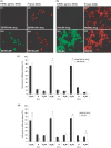
Fig. 1
Fig. 1
BP-induced cell-cycle arrest and apoptosis in GBM cells. DBTRG-05MG and RG2 cells underwent apoptotic cell death after a 48 h treatment with 75 µg/mL BP (a and b), as determined by TUNEL assay for DNA fragmentation (iii and vii) and PI counterstaining for genomic DNA (iv and viii). Control cells were trypsinized and stained in parallel with BP-treated cells (i, ii, v and vi). (c and d) DBTRG-05MG and RG2 cells were arrested in the G0/G1 phase with 75 µg/mL BP. Flow cytometric analysis of DNA contents in the BP-treated (open bars) and control cells (black bars) revealed the proportions of cells at different cell-cycle stages after BP treatment for 6, 12 and 24 h. Each column represents the mean ± SD (*p < 0.05).
Fig. 2
Fig. 2
The expression and activation of apoptosis-associated molecules in GBM cells treated with BP. Whole cell lysates (20 µg/lane) were analyzed with western blotting using specific antibodies to: (a) p53, phospho-p53 and phospho-RB; (b) p16, p21, p27, cdk2, cdk4, cdk6, cyclin D1 and cyclin E; (c) Bax, AIF and caspase 9; (d) Fas, Fas-L, caspase 8 and caspase 3. Lower panel: Relation to control in (a) to (d) is relative to untreated control cells. Positive control of RG2 cells is DBTRG-05 MG cells with BP treatment for 3 h. UD, protein undetectable in this western blot system: ND, not detected.
Fig. 3
Fig. 3
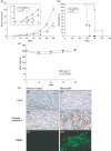
BP inhibition of tumor growth with improved survival rate in a syngenic rat GBM model. RG2 cells (1 × 10⁶) were implanted s.c. into the hind flank region of F344 rats. (a) Tumor size was measured using calipers (*p < 0.05). (b) Survival was monitored daily (p < 0.0001). Rats were killed when tumor size exceeded 25 cm3. Tumor sizes are represented as means ± SEM. There was no statistically significant difference between the control and BP-treated groups with respect to the body weight of rats. (c) Body weights are represented as means ± SEM. (d) Immunohistochemical staining and TUNEL assay were analyzed in GBM tumor tissues (at day 18 after treatment). Representative photographs of sections of the control group (i, iii and v) and BP-treated group (ii, iv and vi) GBM tumors, immunohistochemically stained for cell proliferation marker with Ki-67 (i and ii), cell apoptosis marker with cleaved caspase 3 (iii and iv) and DNA fragmentation of apoptosis cells with TUNEL staining (v and vi). The Ki-67- and caspase 3-positive cells were stained brown and the TUNEL-positive cells were stained green (×400).
Fig. 4
Fig. 4
BP reduction in tumor volume in a syngenic rat GBM tumor in situ model. RG2 cells (5 × 10⁴) were implanted i.c. (striatum) in F344 rats. (a) Tumor volume shown by MRI imaging of serial sections (1.5 μm thick): C1–C6 were from the vehicle control rat; BP1–BP6 from a BP-treated rat (tumor mass shown by white arrow). (b) Tumor volume was calculated using echo-planar imaging capability. Each column represents mean ± SEM (*p < 0.05, **p < 0.001). (c) Immunohistochemical staining and TUNEL assay were performed in rat brain tumor tissues (on day 16 after BP treatment). Representative photographs of sections of the control group (i, iii and v) and BP-treated group (ii, iv and vi) GBM tumors immunohistochemically stained for cell proliferation marker with Ki-67 (i and ii), cell apoptosis marker with caspase 3 (active form; iii and iv) and DNA fragmentation of apoptosis cell with TUNEL staining (v and vi). The Ki-67- and caspase 3-positive cells were stained brown and the TUNEL-positive cells were stained green (×400).
Fig. 5
Fig. 5
BP suppression of xenograft tumor growth of human GBM in nude mice. (a) DBTRG-05MG cells (2.5 × 10⁶) were implanted s.c. into the hind flank region of Foxn1 nu/nu mice. These mice were treated with vehicle as a control (n = 6), BP-70 (70 mg/kg, n = 5), BP-150 (150 mg/kg, n = 5), BP-300 (300 mg/kg, n = 6), BP-500 (500 mg/kg, n = 6) and BP-800 (800 mg/kg, n = 6) s.c. on days 4, 5, 6, 7 and 8. The tumor sizes were measured using calipers. There was a significant difference between the BP-treated group and the control group with respect to the tumor size (p < 0.05). Tumor sizes are presented as mean ± SEM. (b) The survival of mice was monitored daily (p < 0.001). Mice were killed when the tumor size exceeded 1000 mm3. (c) The body weights of mice in the control group, and in the BP-300-, BP-500- and BP-800-treated groups, were not significantly different after BP treatment; body weights are presented as means ± SEM. (d) Immunohistochemical staining and TUNEL assay were performed in human GBM tumor tissues (on day 10 after treatment). Representative photographs of sections of the control group (i, iii and v) and BP-treated group (ii, iv and vi) GBM tumors were immunohistochemically stained for the cell proliferation marker, Ki-67 (i and ii), cell apoptosis marker, caspase 3 (active form; iii and iv), and DNA fragmentation of apoptosis cell with TUNEL staining (v and vi). The Ki-67- and caspase 3-positive cells were stained brown and TUNEL-positive cells were stained green (×400).

References

    1. Ye Y N, Koo M W, Li Y, Matsui H, Cho C H. Angelica sinensis modulates migration and proliferation of gastric epithelial cells. Life Sci. 2001a;68:961–968. - PubMed
    1. Ye Y N, Liu E S, Li Y. Protective effect of polysaccharides-enriched fraction from Angelica sinensis on hepatic injury. Life Sci. 2001b;69:637–646. - PubMed
    1. Graham D I, Lantos P L. Greenfield's Neuropathology. 7. London: Hodder Arnold Press; 2002.
    1. Santarius T, Kirsch M, Rossi M L, Black P M. Molecular aspects of neuro-oncology. ClinNeurolNeurosurg. 1997;99:184–195. - PubMed
    1. Giese A, Bjerkvig R, Berens M E, Westphal M. Cost of migration: invasion of malignant gliomas and implications for treatment. JClinOncol. 2003;21:1624–1636. - PubMed

Publication types

MeSH terms