. 2006 Dec;80(23):11598-609.
doi: 10.1128/JVI.01758-06. Epub 2006 Sep 20.

Characterization of White bream virus reveals a novel genetic cluster of nidoviruses

Affiliations

Characterization of White bream virus reveals a novel genetic cluster of nidoviruses

Heike Schütze et al. J Virol. 2006 Dec.

Abstract

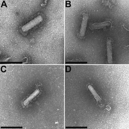
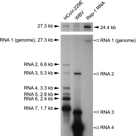
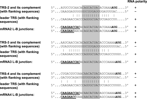
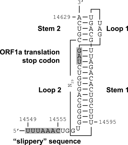
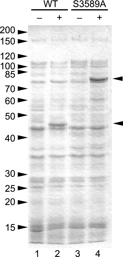
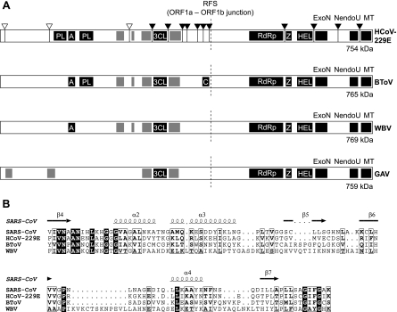
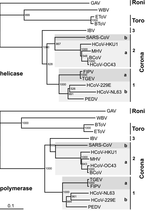
The order Nidovirales comprises viruses from the families Coronaviridae (genera Coronavirus and Torovirus), Roniviridae (genus Okavirus), and Arteriviridae (genus Arterivirus). In this study, we characterized White bream virus (WBV), a bacilliform plus-strand RNA virus isolated from fish. Analysis of the nucleotide sequence, organization, and expression of the 26.6-kb genome provided conclusive evidence for a phylogenetic relationship between WBV and nidoviruses. The polycistronic genome of WBV contains five open reading frames (ORFs), called ORF1a, -1b, -2, -3, and -4. In WBV-infected cells, three subgenomic RNAs expressing the structural proteins S, M, and N were identified. The subgenomic RNAs were revealed to share a 42-nucleotide, 5' leader sequence that is identical to the 5'-terminal genome sequence. The data suggest that a conserved nonanucleotide sequence, CA(G/A)CACUAC, located downstream of the leader and upstream of the structural protein genes acts as the core transcription-regulating sequence element in WBV. Like other nidoviruses with large genomes (>26 kb), WBV encodes in its ORF1b an extensive set of enzymes, including putative polymerase, helicase, ribose methyltransferase, exoribonuclease, and endoribonuclease activities. ORF1a encodes several membrane domains, a putative ADP-ribose 1"-phosphatase, and a chymotrypsin-like serine protease whose activity was established in this study. Comparative sequence analysis revealed that WBV represents a separate cluster of nidoviruses that significantly diverged from toroviruses and, even more, from coronaviruses, roniviruses, and arteriviruses. The study adds to the amazing diversity of nidoviruses and appeals for a more extensive characterization of nonmammalian nidoviruses to better understand the evolution of these largest known RNA viruses.


Figures

FIG. 1.
FIG. 1.
Purified WBV genome RNA is infectious: evidence for viral particle formation and release. The cell culture supernatant from EPC cells transfected with purified WBV genome RNA was analyzed at 6 days posttransfection by electron microscopy (negative staining). (C and D). For comparison, electron micrographs taken from purified WBV virions (27) are shown in panels A and B. Representative pictures of both intact (A and C) and partially opened (B and D) virions are shown. Bar, 150 nm.
FIG. 2.
FIG. 2.
Structural organization and sequence analysis of the WBV (strain DF24/00) genome. (A) Given are the sizes and positions of cDNA clones from WBV genomic libraries that were used to determine the WBV genome sequence. Also shown are the 5′- and 3′-terminal amplicons generated by RACE. (B) Given are the sizes and positions of RT-PCR products used to ascertain the sequence derived from the cDNA clones shown in panel A. (C) Predicted functional ORFs in the WBV genome. Numbers indicate the 5′ and 3′ nucleotides, respectively, of predicted translation start and stop codons. Note that translation of ORF1b is predicted to involve a −1 ribosomal frameshift occurring just upstream of the ORF1a translation stop codon (and downstream of the most 5′-terminal AUG codon that is used here to indicate the ORF1b 5′ end) (for further details, see the text and Table 1). (D) WBV-specific RNAs as determined in this study. The little black box at the 5′ end of the genome indicates the 42-nt leader sequence, which is also present at the 5′ ends of the three subgenome-length RNAs (Fig. 3 and 4). The available evidence from other nidoviruses (37, 47) suggests that attachment of the leader sequence to the coding (body) sequences of WBV subgenome-length RNAs is due to discontinuous extension of subgenome-length minus-strand RNAs. In this process, nascent minus strands switch their template at TRSs located upstream of the S, M, and N genes and bind then to an identical sequence called leader TRS near the 5′ end of the genome, after which the leader sequence is copied to complete negative-strand synthesis (see the text and Fig. 4).
FIG. 3.
FIG. 3.
Detection of WBV genome- and subgenome-length RNAs in virus-infected cells. Northern blot analysis of poly(A)-containing RNA isolated from WBV-infected EPC cells (lane 2). Poly(A) RNAs isolated from HCoV-229E-infected MRC-5 cells (lane 1) and HCoV-229E-derived replicon RNA Rep-1 (lane 3) (28) were used as RNA size markers in this experiment. To detect both the HCoV-229E- and WBV-specific RNAs, a mixture of α-32P-multiprime-labeled probes specific for the 3′-terminal regions of HCoV-229E (nucleotides 26857 to 27277) and WBV (nucleotides 25992 to 26582) was used for hybridization. HCoV-229E genome- and subgenome-length RNAs and the in vitro-transcribed HCoV-229E Rep-1 RNA are indicated by black arrowheads, with sizes given in kilobases. White arrowheads indicate the four WBV-specific RNAs detected in this experiment. The longer exposure presented above shows the size of the WBV genomic RNA more clearly and allows its size to be compared with those of the 27.3- and 24.4-kb marker RNAs. The calculated sizes (Table 1 and Fig. 4) of the sg RNAs are 5,162 nts (RNA 2), 1,475 nts (RNA 3), and 774 nts (RNA 4) [including the 5′ leader but excluding the poly(A) tail].
FIG. 4.
FIG. 4.
Leader-body junctions in WBV subgenomic RNAs. Shown are the junction sites between a short sequence, called leader, that is derived from the 5′ end of the genome, and the coding (so-called body) sequences of subgenomic RNAs 2 to 4. The gray box highlights a sequence, CA(G/A)CACUAC, and its negative-strand complement that we predict to act as a core TRS element in WBV. As in corona- and arteriviruses (37, 47), this leader sequence is conserved near the 5′ end of the genome (nts 44 to 52) and upstream of the translation start codon of each of the downstream ORFs specifying the viral structural proteins, S, M, and N. For each of the core TRS elements, the flanking sequences in the WBV genome RNA and the corresponding minus-strand sequence are given. Possible base-pairing interactions between the minus strand and the proposed leader TRS are indicated, and the leader-body (L-B) junction in the respective mRNA, as determined by RT-PCR and sequencing, is given below. Sequences derived from the 5′ end of the genome (leader sequence) are boldfaced and underlined. Translation start codons of the S (RNA 2), M (RNA 3), and N (RNA 4) genes are boldfaced and italicized. Please note that, with respect to the minus-strand sequence, the actual fusion appears to occur slightly downstream of the fully complementary sequence rather than within this particular sequence.
FIG. 5.
FIG. 5.
Model of the WBV ribosomal frameshifting element. By analogy with other nidoviruses, the element is proposed to consist of a putative RNA pseudoknot structure (comprised of two stems and two loops) and a slippery sequence (14549UUUAAAC15555) at which the actual frameshift is predicted to occur. The sequences boxed in gray indicate the predicted slippery sequence and the ORF1a translation termination codon.
FIG. 6.
FIG. 6.
Proteolytic activity of WBV pp1a/pp1ab amino acid residues Ser3424 toGln3726. Total cell lysates from E. coli TB1 cells transformed with pMal-WBV-3CL_559-560 (WT) (lanes 1 and 2) and pMal-WBV-3CL_S3589A (S3589A) (lanes 3 and 4) were separated by sodium dodecyl sulfate-polyacrylamide gel electrophoresis in a 12.5% polyacrylamide gel and stained with Coomassie brilliant blue R-250. The bacteria were mock induced (lanes 1 and 3) or induced with 1 mM IPTG for 3 h (lanes 2 and 4). The positions of the fusion protein and cleavage product are indicated by arrowheads. The molecular masses of marker proteins are given in kDa to the left.
FIG. 7.
FIG. 7.
Domain organization of nidovirus replicase polyproteins. (A) Comparison of the WBV pp1ab domain organization with those of representative viruses from other nidovirus genera: HCoV-229E (genus Coronavirus and family Coronaviridae), bovine torovirus (BToV; genus Torovirus and family Coronaviridae), and gill-associated virus (GAV; genus Okavirus and family Roniviridae). The polyproteins are processed by viral proteases that are part of the polyprotein. The coronavirus pp1ab proteolytic processing has been characterized in considerable detail (71, 75) and is illustrated here for HCoV-229E. To produce a total of 16 nonstructural proteins, three cleavages are carried out by papain-like proteases (PL) in the N-proximal region of the polyprotein (indicated by white arrowheads), and 11 cleavages are carried out by the 3C-like protease (3CL) in the central and C-terminal parts of the polyprotein (indicated by black arrowheads). For the genera Torovirus and Okavirus, only limited information on proteases and their cleavage sites is available (53, 72). The putative proteases of BToV have not been characterized, and only a few 3C-like protease cleavage sites (not shown) have been identified for GAV (72). Proteases and other conserved enzymatic activities are indicated by black boxes. A, ADP-ribose 1"-phosphatase (ADRP) related to cellular macro domain proteins (20, 40); Z, zinc-binding domain (51); HEL, helicase domain (50); ExoN, 3′-to-5′ exoribonuclease (34); MT, putative ribose-2′-O-methyltransferase domain (21, 54); C, putative cyclic nucleotide phosphodiesterase (54); RFS, ribosomal frameshift site. Regions with predicted transmembrane domains (see Materials and Methods) are indicated by gray boxes. Note that the expression of the C-terminal part of pp1ab requires a ribosomal frameshift into ORF1b, which is predicted to occur just upstream of the ORF1a translation stop codon (Fig. 5). The sizes and positions of the polyproteins and functional domains are not precisely drawn to scale. (B) Partial sequence alignment of ADRP domains from SARS-CoV and HCoV-229E, whose activities have been characterized previously (20, 40, 41, 43), and the predicted ADRP domains from BToV (18) and WBV (this study). The alignment was generated using the ClustalX program (version 1.8). The secondary structure information was derived from the published SARS-CoV ADRP crystal structure (Protein Data Bank no. 2ACF) (43) and, together with the alignment, used as input for the ESPript program, version 2.2 (http://prodes.toulouse.inra.fr/ESPript/cgi-bin/ESPript.cgi). Sequences of the proteins were derived from the DDBJ/EMBL/GenBank database accession numbers NC_002645 (HCoV-229E pp1a/pp1ab residues Phe1299 to Lys1398), AY291315 (SARS-CoV pp1a/pp1ab residues Val1034 to Lsy1135), AY427798 (BToV [strain Breda-1] pp1a/pp1ab residues Y1668 to Ser1775), and DQ898157 (WBV pp1a/pp1ab residues Phe1667 to Lys1796). Black boxes, identical residues; white boxes, similar residues.
FIG. 8.
FIG. 8.
Phylogenetic analysis of WBV helicase and polymerase core domains. Phylogenetic trees were generated from multiple-sequence alignments of the most conserved regions of nidovirus RNA-dependent RNA polymerase (residues Thr4723 to Gln5396 in the WBV pp1ab sequence) and helicase domains (residues Ala5644 to Cys5924 in the WBV pp1ab sequence), using the neighbor-joining algorithm as implemented in the ClustalX 1.8 program (for details, see Materials and Methods). The WBV sequences were compared with those from Gill-associated virus (GAV, accession no. AF227196), Equine torovirus Berne (EToV, X52374), and Bovine torovirus Breda-1 (BToV, AY427798) as well as from viruses representing the three coronavirus groups, including the recently introduced subgroups 1a, 1b, 2a, and 2b (26). Group 1a, Transmissible gastroenteritis virus Purdue-115 (TGEV, accession no. Z34093) and Feline infectious peritonitis virus WSU 79/1146 (FIPV, DQ010921); group 1b, HCoV-229E (NC_002645), Human coronavirus NL63 Amsterdam I (HCoV-NL63, AY567487), and Porcine epidemic diarrhea virus CV777 (PEDV, AF353511); group 2a, Bovine coronavirus LUN (BCoV, AF391542), Human coronavirus OC43 serotype Paris (HCoV-OC43, AY585229), Mouse hepatitis virus A59 (MHV, NC_001846), and Human coronavirus HKU1 (HCoV-HKU1, NC_006577); group 2b, Severe acute respiratory syndrome coronavirus Frankfurt 1 (SARS-CoV, AY291315); group 3, Avian infectious bronchitis virus Beaudette (IBV, NC_001451).

Similar articles

Cited by

References

    1. Anand, K., G. J. Palm, J. R. Mesters, S. G. Siddell, J. Ziebuhr, and R. Hilgenfeld. 2002. Structure of coronavirus main proteinase reveals combination of a chymotrypsin fold with an extra alpha-helical domain. EMBO J. 21:3213-3224. - PMC - PubMed
    1. Anand, K., J. Ziebuhr, P. Wadhwani, J. R. Mesters, and R. Hilgenfeld. 2003. Coronavirus main proteinase (3CLpro) structure: basis for design of anti-SARS drugs. Science 300:1763-1767. - PubMed
    1. Barrette-Ng, I. H., K. K. Ng, B. L. Mark, D. Van Aken, M. M. Cherney, C. Garen, Y. Kolodenko, A. E. Gorbalenya, E. J. Snijder, and M. N. James. 2002. Structure of arterivirus nsp4. The smallest chymotrypsin-like proteinase with an alpha/beta C-terminal extension and alternate conformations of the oxyanion hole. J. Biol. Chem. 277:39960-39966. - PubMed
    1. Bartlam, M., H. Yang, and Z. Rao. 2005. Structural insights into SARS coronavirus proteins. Curr. Opin. Struct. Biol. 15:664-672. - PMC - PubMed
    1. Bendtsen, J. D., H. Nielsen, G. von Heijne, and S. Brunak. 2004. Improved prediction of signal peptides: SignalP 3.0. J. Mol. Biol. 340:783-795. - PubMed

Publication types

MeSH terms

LinkOut - more resources