. 2006 Dec;80(23):11539-55.
doi: 10.1128/JVI.01016-06. Epub 2006 Sep 20.

Human cytomegalovirus (HCMV) infection of endothelial cells promotes naive monocyte extravasation and transfer of productive virus to enhance hematogenous dissemination of HCMV

Affiliations

Human cytomegalovirus (HCMV) infection of endothelial cells promotes naive monocyte extravasation and transfer of productive virus to enhance hematogenous dissemination of HCMV

Gretchen L Bentz et al. J Virol. 2006 Dec.

Abstract

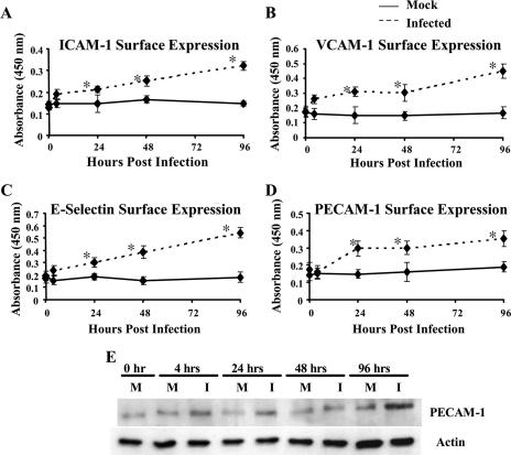
Human cytomegalovirus (HCMV) pathogenesis is dependent on the hematogenous spread of the virus to host tissue. While data suggest that infected monocytes are required for viral dissemination from the blood to the host organs, infected endothelial cells are also thought to contribute to this key step in viral pathogenesis. We show here that HCMV infection of endothelial cells increased the recruitment and transendothelial migration of monocytes. Infection of endothelial cells promoted the increased surface expression of cell adhesion molecules (intercellular cell adhesion molecule 1, vascular cell adhesion molecule 1, E-selectin, and platelet endothelial cell adhesion molecule 1), which were necessary for the recruitment of naïve monocytes to the apical surface of the endothelium and for the migration of these monocytes through the endothelial cell layer. As a mechanism to account for the increased monocyte migration, we showed that HCMV infection of endothelial cells increased the permeability of the endothelium. The cellular changes contributing to the increased permeability and increased naïve monocyte transendothelial migration include the disruption of actin stress fiber formation and the decreased expression of lateral junction proteins (occludin and vascular endothelial cadherin). Finally, we showed that the migrating monocytes were productively infected with the virus, documenting that the virus was transferred to the migrating monocyte during passage through the lateral junctions. Together, our results provide evidence for an active role of the infected endothelium in HCMV dissemination and pathogenesis.


Figures

FIG. 1.
FIG. 1.
In vitro model for studying the effects of viral infection on monocyte diapedesis. HMECS were grown to confluence on polystyrene transwell plates and infected with HCMV (MOI, 20), mock infected, or treated with PMA (10 ng/ml) or LPS (10 μg/ml). After 24 h, the endothelial cells were washed and monocytes labeled with a green cell tracker dye were added to the wells. At 24 h after the addition of monocytes, three random FOV of monocytes on the HMECS in the top of the insert were counted to determine the percentage of cells adhering to the endothelial cell layer and beginning to undergo diapedesis versus the percentage of cells rounded up and stationary on the endothelial cell layer. At 96 h after the addition of monocytes, the number of monocytes that migrated completely through the endothelial cell layer was determined for 10 random FOV in the bottom chamber. The percentage of added monocytes that migrated through the wells was also determined at 96 h after the addition of monocytes to the transwells.
FIG. 2.
FIG. 2.
HCMV infection of endothelial cells promotes naïve monocyte recruitment and transendothelial migration. (A) HCMV infection of endothelial cells resulted in increased naïve monocyte recruitment. HMECs were mock infected, HCMV infected (MOI, 20), PMA treated (10 ng/ml), or LPS treated (10 μg/ml) for 24 h. Human monocytes were isolated, labeled with a green cell tracker dye, and added to each well. At 24 h after the addition of monocytes, the percentage of monocytes undergoing diapedesis versus the percentage of monocytes rounded up and stationary on the endothelial cell layer was determined. Results are plotted as means ± SD of three random fields of view. Magnification, ×200. (B) HCMV infection of endothelial cells resulted in increased naïve monocyte transendothelial migration. At 96 h after the addition of monocytes, the number of monocytes that migrated completely through the endothelial cell layer was determined. Results are plotted as means ± SD of 10 random fields of view. Magnification, ×200. (C) HCMV infection of endothelial cells resulted in increased total naïve monocyte transendothelial migration. At 96 h after the addition of monocytes to the transwell inserts, monocytes that had migrated through both the endothelial cell layer and the insert and had adhered to the bottom of the well were collected and counted. The number of monocytes that migrated through the endothelium and the transwell versus the total number of monocytes added to the wells was determined. Results are plotted as means ± SD of the percentage of added monocytes that underwent transendothelial migration. Results are an average of four independent experiments from separate human blood donors. HCMV-infected, LPS-treated, and PMA-treated groups were significantly different (P < 0.01) than the mock-infected group.
FIG. 3.
FIG. 3.
HCMV infection of endothelial cells resulted in the upregulation of cell adhesion molecule expression. (A to D) HCMV infection of endothelial cells promoted increased surface expression of cell adhesion molecules. Cell-based ELISAs were performed as previously described (78) to examine the surface expression of ICAM-1 (A), VCAM-1 (B), E-selectin (C), and PECAM-1 (D) at the indicated times after infection. The straight line corresponds to mock-infected cells, while the dashed line corresponds to HCMV-infected cells (MOI, 20). Results are plotted as means ± SD of three independent experiments performed in triplicate. HCMV-infected HMECs showed a significant increase (P < 0.01) in surface expression of ICAM-1, VCAM-1, E-selectin, and PECAM-1 by 24 hpi compared to mock-infected HMECs. (E) HCMV infection of endothelial cells promoted increased PECAM-1 protein expression. Western blot analyses of PECAM-1 and actin were performed using equal protein loading of mock-infected and HCMV-infected (MOI, 20) HMEC lysates harvested at the indicated times after infection.
FIG. 4.
FIG. 4.
HCMV-induced cell adhesion molecule surface expression is functional. (A) The increased surface expression of ICAM-1, VCAM-1, and E-selectin following HCMV infection is necessary for the recruitment of naïve monocytes. HMECs were mock infected or HCMV infected (MOI, 20) for 24 h. Cells were washed and incubated for 1 h at 37°C with medium containing blocking antibodies (ICAM-1, 20 μg/ml; VCAM-1, 30 μg/ml; E-selectin, 50 μg/ml; all three antibodies; or the IgG1 isotype control, 50 μg/ml), after which the cells were washed again and isolated labeled human monocytes were added to each well. At 24 h after the addition of monocytes, the percentage of monocytes undergoing diapedesis versus the percentage of monocytes rounded up and stationary on the endothelial cell layer was determined. Results are plotted as means ± SD of three random fields of view. Magnification, ×200. (B) Blocking the adhesion of monocytes to the HCMV-infected endothelial cells inhibited HCMV-induced naïve monocyte transendothelial migration. At 96 h after the addition of monocytes, the number of monocytes that migrated completely through the endothelial cell layer was determined. Results are plotted as means ± SD of 10 random fields of view. Magnification, ×200.
FIG. 5.
FIG. 5.
HCMV infection of endothelial cells resulted in increased endothelial cell permeability. (A) Diffusion of a TBA complex (68) across a confluent monolayer of mock-infected, HCMV-infected (MOI, 20), or cytochalasin D-treated (1 μM) HMECS or across the insert alone was determined at the indicated times. Results are plotted as means ± SD of three independent experiments performed in triplicate. (B) TER of a confluent monolayer of mock-infected, HCMV-infected (MOI, 20), or cytochalasin D-treated (1 μM) HMECS or across the insert alone was determined at various times after treatment. Results are plotted as means ± SD of three independent experiments done in triplicate. HCMV-infected and cytochalasin D-treated HMECs showed a significant increase (P < 0.01) in permeability compared to mock-infected HMECs.
FIG. 6.
FIG. 6.
HCMV infection of endothelial cells resulted in decreased junctional protein expression. (A) Western blot analyses of actin, VE-cadherin, and occludin protein levels were performed using equal protein loading of mock-infected and HCMV-infected (MOI, 20) HMEC lysates harvested at the indicated times after infection. (B and C) Bands were analyzed by densitometry to determine relative levels of occludin (both the 65- and 72-kDa forms) and VE-cadherin (130 kDa) (B) and the alternative forms of VE-cadherin (lower molecular mass, ∼100 kDa; represented as lower MW VE-cadherin) (C) and are expressed in arbitrary units as a ratio of x/actin. A representative experiment from three independent experiments is shown.
FIG. 7.
FIG. 7.
HCMV infection of endothelial cells promoted junctional protein internalization. HMECs were grown to confluence on fibronectin-coated coverslips and mock infected or HCMV infected (MOI, 20) for 96 h. The cells were then fixed, stained with the appropriate primary and secondary antibodies and DAPI, and examined by immunofluorescence microscopy. Magnification, ×1,000. (A and B) Expression of occludin (A) and VE-cadherin (B) was examined in mock-infected cells. (C and D) Using the same exposure time, expression of occludin (C) and VE-cadherin (D) was examined in HCMV-infected cells. (E and F) A longer exposure time was used to examine the expression of occludin (E) and VE-cadherin (F) in HCMV-infected cells. A representative experiment from three independent experiments is shown.
FIG. 8.
FIG. 8.
HCMV-induced lateral junction disruption promotes increased endothelial cell permeability and naïve monocyte transendothelial migration. (A) TER of a confluent monolayer of endothelial cells treated with a VE-cadherin-specific blocking antibody (cl75; 25 μg/ml) or the isotype control antibody (50 μg/ml) and either mock infected, HCMV infected (MOI, 20), or cytochalasin D treated (1 μM) was determined at various times after treatment. The resistance across the empty insert was also determined. Results are plotted as means ± SD of three independent experiments performed in triplicate. HMECs that were infected or infected and treated with the isotype control, with the VE-cadherin-specific blocking antibody, or the IgG1 isotype control and mock infected or HCMV infected or treated with cytochalasin D showed a significant increase (P < 0.01) in permeability compared to mock-infected HMECs. (B) Blocking VE-cadherin-based cell adhesion promotes naïve monocyte transendothelial migration. HMECs were mock infected or HCMV infected (MOI, 20) for 24 h. Cells were washed, and medium containing the VE-cadherin-specific blocking antibody (25 μg/ml) or the IgG1 isotype control (50 μg/ml) was added to each well. Isolated and labeled human monocytes were added to each well. At 96 h after the addition of monocytes, the number of monocytes that migrated completely through the endothelial cell layer was determined. Results are plotted as means ± SD of 10 random fields of view. Magnification, ×200.
FIG. 9.
FIG. 9.
HCMV infection of endothelial cells disrupts actin stress fiber formation. HMECs were grown on fibronectin-coated coverslips and mock infected or HCMV infected (MOI, 20) for 24 h. The cells were then fixed, stained with phalloidin conjugated to Alexa Fluor 488 (A and B) or 546 (C and D) and DAPI (C and D), and examined by immunofluorescence microscopy. Stress fiber formation in mock-infected and HCMV-infected HMECs was examined at ×400 (A) and ×1,000 (B) magnification. Representative images of two different experiments from a total of three independent experiments are shown.
FIG. 10.
FIG. 10.
HCMV can be transferred from the infected endothelial cell to a monocyte undergoing transendothelial migration. HMECs were grown to confluence in tissue culture inserts and mock infected or HCMV infected (Towne/E or TB40-UL32-HCMV/E; MOI, 20) for 24 h. Isolated peripheral blood monocytes were then added to the wells. At 96 h after the addition of monocytes, cells that migrated through the insert into the bottom of the well were collected on the fibronectin-coated coverslips in the bottom of the well and fixed and permeabilized. The cells were then stained with TO-PRO3-iodide (A and B) or DAPI (C and D) and an anti-pp65 antibody or an anti-GFP antibody and examined by immunofluorescence microscopy. (A and B) Confocal microscopy was performed to examine the transfer of pp65 from the infected endothelial cells to the migrating naïve monocytes. Only monocytes that migrated through the HCMV-infected endothelial cells stained positively for pp65. Magnification, ×1,000. (C and D) Bright-field and immunofluorescence microscopy was performed to examine the transfer of HCMV from the infected endothelial cells to migrating naïve monocytes. Only monocytes that migrated through the HCMV-infected endothelial cells stained positively for TB40-UL32-GFP HCMV. Magnification, ×1,000. (E) A PCR/Southern blot analysis was utilized to confirm the presence of HCMV genomic DNA in various populations of monocytes. Lane 1, monocytes that migrated through mock-infected HMECs; lane 2, monocytes that migrated through TB40-UL32-GFP HCMV-infected HMECs; lane 3, mock-infected monocytes; lane 4, HCMV-infected monocytes; lane 5, water control. Only monocytes that migrated through HCMV-infected HMECs or monocytes that were directly infected with HCMV were positive for HCMV genomic DNA, suggesting that viral DNA can be transferred from the infected endothelial cell to the migrating, naïve monocyte.

References

    1. Adam, E., J. L. Melnick, J. L. Probtsfield, B. L. Petrie, J. Burek, K. R. Bailey, C. H. McCollum, and M. E. DeBakey. 1987. High levels of cytomegalovirus antibody in patients requiring vascular surgery for atherosclerosis. Lancet ii:291-293. - PubMed
    1. Albelda, S. M., W. A. Muller, C. A. Buck, and P. J. Newman. 1991. Molecular and cellular properties of PECAM-1 (endoCAM/CD31): a novel vascular cell-cell adhesion molecule. J. Cell Biol. 114:1059-1068. - PMC - PubMed
    1. Allport, J. R., W. A. Muller, and F. W. Luscinskas. 2000. Monocytes induce reversible focal changes in vascular endothelial cadherin complex during transendothelial migration under flow. J. Cell Biol. 148:203-216. - PMC - PubMed
    1. Bale, J. F., Jr., and M. E. O'Neil. 1989. Detection of murine cytomegalovirus DNA in circulating leukocytes harvested during acute infection of mice. J. Virol. 63:2667-2673. - PMC - PubMed
    1. Berman, M. E., Y. Xie, and W. A. Muller. 1996. Roles of platelet/endothelial cell adhesion molecule-1 (PECAM-1, CD31) in natural killer cell transendothelial migration and beta 2 integrin activation. J. Immunol. 156:1515-1524. - PubMed

Publication types

MeSH terms