. 2006 Dec;80(23):11467-74.
doi: 10.1128/JVI.01125-06. Epub 2006 Sep 20.

Crystal structure of the West Nile virus envelope glycoprotein

Affiliations

Crystal structure of the West Nile virus envelope glycoprotein

Grant E Nybakken et al. J Virol. 2006 Dec.

Abstract

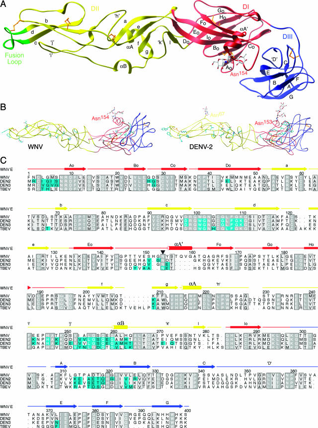
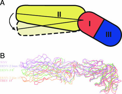
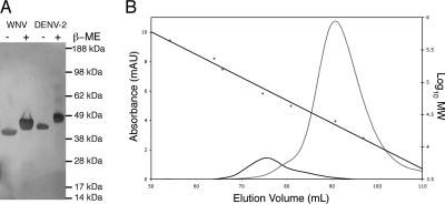
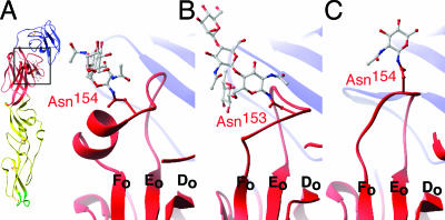
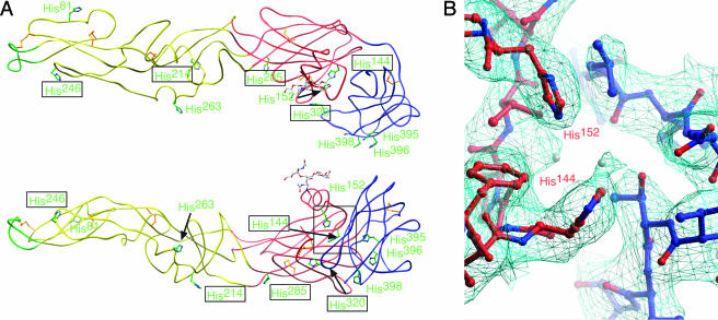
The envelope glycoprotein (E) of West Nile virus (WNV) undergoes a conformational rearrangement triggered by low pH that results in a class II fusion event required for viral entry. Herein we present the 3.0-A crystal structure of the ectodomain of WNV E, which reveals insights into the flavivirus life cycle. We found that WNV E adopts a three-domain architecture that is shared by the E proteins from dengue and tick-borne encephalitis viruses and forms a rod-shaped configuration similar to that observed in immature flavivirus particles. Interestingly, the single N-linked glycosylation site on WNV E is displaced by a novel alpha-helix, which could potentially alter lectin-mediated attachment. The localization of histidines within the hinge regions of E implicates these residues in pH-induced conformational transitions. Most strikingly, the WNV E ectodomain crystallized as a monomer, in contrast to other flavivirus E proteins, which have crystallized as antiparallel dimers. WNV E assembles in a crystalline lattice of perpendicular molecules, with the fusion loop of one E protein buried in a hydrophobic pocket at the DI-DIII interface of another. Dimeric E proteins pack their fusion loops into analogous pockets at the dimer interface. We speculate that E proteins could pivot around the fusion loop-pocket junction, allowing virion conformational transitions while minimizing fusion loop exposure.


Figures

FIG.< 1.
FIG.< 1.
Structure of WNV E protein. (A) DI (red), DII (yellow), and DIII (blue) of monomeric E adopt a topology typical of E proteins from other flaviviruses. The fusion loop (residues 98 to 110) is highlighted in green at the tip of DII. The carbohydrate on Asn154, colored according to atomicity, of DI protrudes next to a novel α-helix. Disulfide bonds are shown in gold. (B) (Left) Tube representation of WNV E, with potential dimer contacts shown in cyan. (Right) Tube representation of DENV-2 E (1OAN), with known dimer contacts shown in cyan. (C) Structural alignment of E structures from WNV, DENV-2, DENV-3, and TBEV. Contact residues within 4.0 Å of a dimer mate are colored cyan. Conservation and similarity are depicted in dark and light gray, respectively. Note the high degree of conservation among the dimerization residues. The glycosylation site is marked with an inverted triangle.
FIG. 2.
FIG. 2.
WNV E has a unique interdomain orientation most similar to the structure of immature E. (A) Schematic showing the angle that was measured for each orientation of the DI-DII angle. (B) C-α traces of the WNV, DENV-3 (1UZG), and TBEV (1SVB) mature E crystal structures and the immature (1TGE) and mature (1P58) DENV-2 cryo-EM reconstructions. They overlap in DI, revealing the variability in DI-DII orientations.
FIG. 3.
FIG. 3.
DENV-2 E favors dimers more than WNV E does. (A) Silver-stained gel demonstrating similar molecular masses for DENV-2 E (calculated molecular mass, 46 kDa) and WNV E (calculated molecular mass, 44 kDa). Note that DENV-2 E has an additional glycosylation site. (B) Size-exclusion profiles for WNV E (gray) and DENV-2 E (black). The straight black line shows the calibration of the column (right axis). WNV E eluted with an observed molecular mass of 22 kDa, and DENV-2 E eluted with an observed molecular mass of 77 kDa.
FIG. 4.
FIG. 4.
The fusion loop of the WNV E monomer packs in the same pocket as that used by other flavivirus E dimers. (A) Scheme demonstrating the relative orientations of the E proteins for fusion loop packing in DENV-2 (left) and WNV (right). (B) The green worm of the fusion loop packs against similar portions of DI and DIII (magenta) in both WNV (right) and DENV-2 (left). Equivalent contact residues in both structures are labeled in black, and other contacts are shown in white.
FIG. 5.
FIG. 5.
Glycosylation of WNV E is shifted by a unique α-helix. (A) Ribbon figure of WNV E demonstrating a boxed area that is enlarged and shows the glycosylation (gray) of WNV E situated closely apposed to the αA′ helix. (B) Glycosylation of DENV-2 E (1OAN). (C) Glycosylation of tick-borne encephalitis virus E (1SVB).
FIG. 6.
FIG. 6.
Histidines are located at hinge regions of flavivirus E proteins. (A) Tube representation of WNV E, with histidine residues depicted in green, viewed from outside the virion (top) and from the perspective of a dimer partner (bottom) inside the virion. Histidines with boxed labels are structurally conserved between flavivirus structures. (B) Detailed depiction of the electron density (cyan) surrounding the histidine residues forming a base for DIII. Water molecules are depicted as gray spheres.

References

    1. Allison, S. L., J. Schalich, K. Stiasny, C. W. Mandl, and F. X. Heinz. 2001. Mutational evidence for an internal fusion peptide in flavivirus envelope protein E. J. Virol. 75:4268-4275. - PMC - PubMed
    1. Beasley, D. W., and A. D. Barrett. 2002. Identification of neutralizing epitopes within structural domain III of the West Nile virus envelope protein. J. Virol. 76:13097-13100. - PMC - PubMed
    1. Beasley, D. W., C. T. Davis, J. Estrada-Franco, R. Navarro-Lopez, A. Campomanes-Cortes, R. B. Tesh, S. C. Weaver, and A. D. Barrett. 2004. Genome sequence and attenuating mutations in West Nile virus isolate from Mexico. Emerg. Infect. Dis. 10:2221-2224. - PMC - PubMed
    1. Beasley, D. W., M. C. Whiteman, S. Zhang, C. Y. Huang, B. S. Schneider, D. R. Smith, G. D. Gromowski, S. Higgs, R. M. Kinney, and A. D. Barrett. 2005. Envelope protein glycosylation status influences mouse neuroinvasion phenotype of genetic lineage 1 West Nile virus strains. J. Virol. 79:8339-8347. - PMC - PubMed
    1. Bressanelli, S., K. Stiasny, S. L. Allison, E. A. Stura, S. Duquerroy, J. Lescar, F. X. Heinz, and F. A. Rey. 2004. Structure of a flavivirus envelope glycoprotein in its low-pH-induced membrane fusion conformation. EMBO J. 23:728-738. - PMC - PubMed

Publication types

Substances

Associated data

LinkOut - more resources