Comparative Study
. 2006 Sep 20;26(38):9646-55.
doi: 10.1523/JNEUROSCI.1620-06.2006.

Schwann cells express motor and sensory phenotypes that regulate axon regeneration

Affiliations
Comparative Study

Schwann cells express motor and sensory phenotypes that regulate axon regeneration

A Höke et al. J Neurosci. .

Abstract

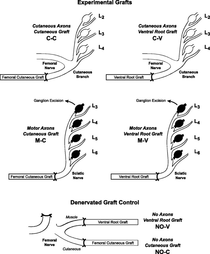
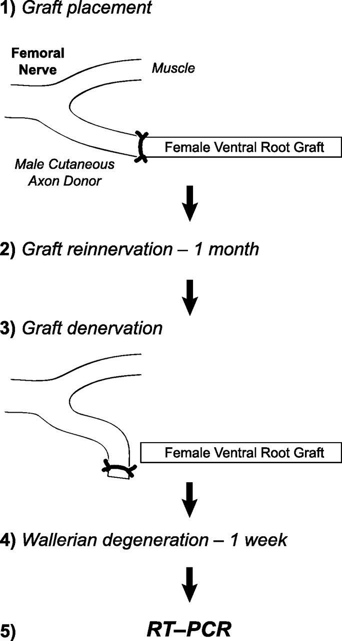
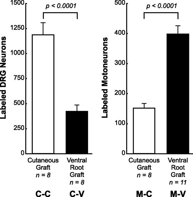
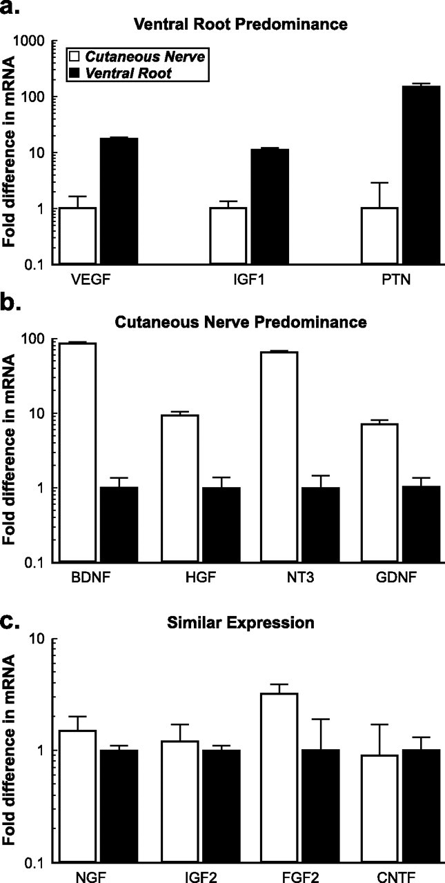
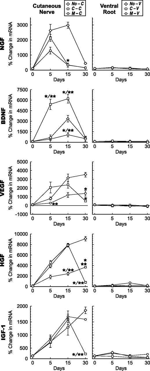
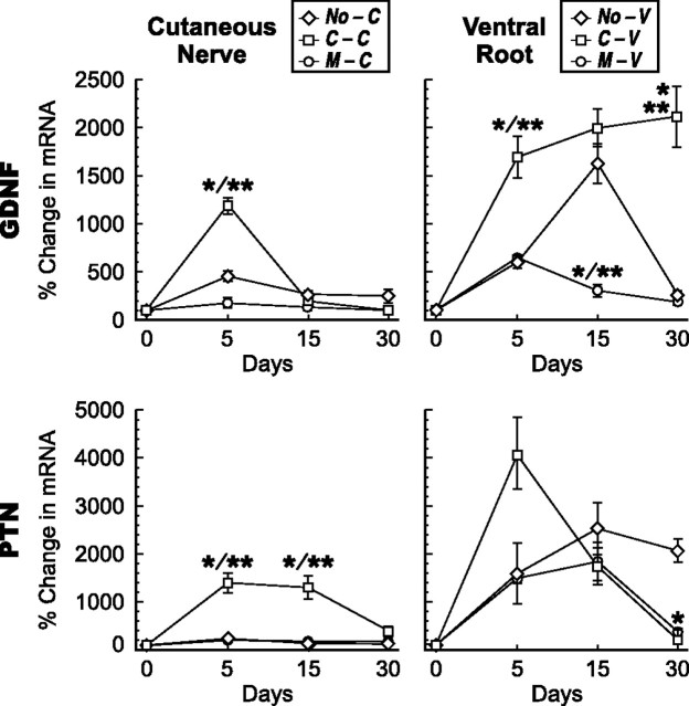
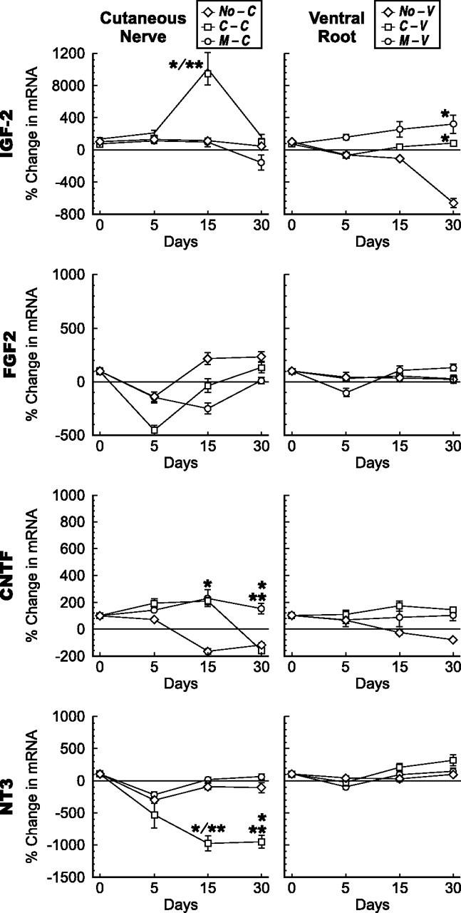
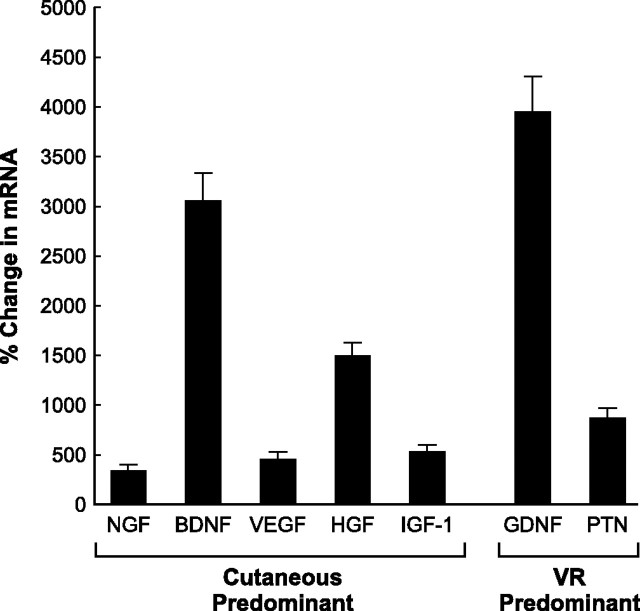
Schwann cell phenotype is classified as either myelinating or nonmyelinating. Additional phenotypic specialization is suggested, however, by the preferential reinnervation of muscle pathways by motoneurons. To explore potential differences in growth factor expression between sensory and motor nerve, grafts of cutaneous nerve or ventral root were denervated, reinnervated with cutaneous axons, or reinnervated with motor axons. Competitive reverse transcription-PCR was performed on normal cutaneous nerve and ventral root and on graft preparations 5, 15, and 30 d after surgery. mRNA for nerve growth factor (NGF), brain-derived neurotrophic factor (BDNF), vascular endothelial growth factor, hepatocyte growth factor, and insulin-like growth factor-1 was expressed vigorously by denervated and reinnervated cutaneous nerve but minimally by ventral root. In contrast, mRNA for pleiotrophin (PTN) and glial cell line-derived neurotrophic factor was upregulated to a greater degree in ventral root. ELISA confirmed that NGF and BDNF protein were significantly more abundant in denervated cutaneous nerve than in denervated ventral root, but that PTN protein was more abundant in denervated ventral root. The motor phenotype was not immutable and could be modified toward the sensory phenotype by prolonged reinnervation of ventral root by cutaneous axons. Retrograde labeling to quantify regenerating neurons demonstrated that cutaneous nerve preferentially supported cutaneous axon regeneration, whereas ventral root preferentially supported motor axon regeneration. Schwann cells thus express distinct sensory and motor phenotypes that are associated with the support of regeneration in a phenotype-specific manner. These findings suggest that current techniques of bridging gaps in motor and mixed nerve with cutaneous graft could be improved by matching axon and Schwann cell properties.


Figures

Figure 1.
Figure 1.
Surgical preparations analyzed in these experiments. The cutaneous branch of the femoral nerve was used as a source of cutaneous axons and the sciatic nerve, deafferented by removal of dorsal root ganglia L3, L4, L5, and L6, as a pure source of motor axons. Grafts were obtained from the femoral cutaneous nerve or the L4 or L5 ventral roots. Experimental groups are identified as to axon source (C, cutaneous axons; M, motor axons) and as to graft type (C, cutaneous nerve graft; V, ventral root graft); the label C-V thus identifies preparations in which cutaneous axons grow into ventral root grafts. Denervated graft controls were provided by suturing cutaneous nerve and ventral root grafts to the femoral cutaneous and muscle branches and then interrupting the donor femoral nerve proximally during the same procedure to allow normal graft revascularization while preventing axon ingrowth. Because no axons are present in these controls, the letters NO identify the axon source (e.g., NO-V describes preparations in which ventral root grafts are revascularized without the possibility of axon ingrowth).
Figure 2.
Figure 2.
Phenotype plasticity experiment. Ventral root grafts harvested from female Lewis rats were sewn to the femoral cutaneous branch of male Lewis rats. After 1 month of inappropriate reinnervation by cutaneous axons, the grafts were denervated for 1 week and their expression profiles were determined by RT-PCR. A portion of the proximal graft was excised at the time of denervation to exclude tissue that could have been invaded by host Schwann cells. PCR for a Y-chromosome-specific gene was performed on genomic DNA isolated from the grafts to ensure that the remaining Schwann cells were of graft origin.
Figure 3.
Figure 3.
Results of retrograde labeling in the four experimental groups, identified as to axon source and graft type as in Figure 1. Label was applied to the center of the graft after reinnervation had progressed for 2 weeks. Labeled neurons were then counted in the spinal cord or L2, L3, and L4 DRGs as appropriate. Sensory neuron regeneration was preferentially supported by cutaneous nerve grafts, whereas motor axon regeneration was preferentially supported by ventral root grafts.
Figure 4.
Figure 4.
Comparison of baseline growth factor expression in intact ventral root and cutaneous nerve. Each factor is characterized as to whether relative mRNA levels were higher in ventral root (a), higher in cutaneous nerve (b), or similar in both types of nerve (c). For each comparison, the lower expression level is normalized as 1 arbitrary unit of expression.
Figure 5.
Figure 5.
Growth factors expressed predominately in cutaneous nerve. Changes in mRNA levels in cutaneous nerve grafts (left column) and ventral root grafts (right column) after denervation, and reinnervation by motor or sensory axons at 5, 15, and 30 d. The abbreviations established in Figure 1 are used to identify the groups. *p < 0.005 compared with denervation alone; **p < 0.005 motor versus sensory reinnervation.
Figure 6.
Figure 6.
Growth factors expressed predominately in ventral root. Changes in mRNA levels in cutaneous nerve grafts (left column) and ventral root grafts (right column) after denervation, and reinnervation by motor or sensory axons at 5, 15, and 30 d. The abbreviations established in Figure 1 are used to identify the groups. *p < 0.005 compared with denervation alone; **p < 0.005 motor versus sensory reinnervation.
Figure 7.
Figure 7.
Growth factors that lack modality specificity. Changes in mRNA levels in cutaneous nerve graft (left column) and ventral root graft (right column) after denervation, and reinnervation by motor or sensory axons at 5, 15, and 30 d. The abbreviations established in Figure 1 are used to identify the groups. *p < 0.005 compared with denervation alone; **p < 0.005 motor versus sensory reinnervation.
Figure 8.
Figure 8.
ELISA for NGF, GDNF, PTN, and BDNF was performed on protein extracted from normal cutaneous nerve (C-0), normal ventral root (V-0), denervated cutaneous nerve (C-15), and denervated ventral root (V-15). *p < 0.05; **p < 0.005. Protein levels confirm selective upregulation of NGF and BDNF in denervated cutaneous nerve and PTN in denervated ventral root. Error bars represent 1 SD from the mean.
Figure 9.
Figure 9.
Results of phenotypic plasticity experiment (Fig. 2). Changes in mRNA levels of ventral root grafts that were transected after 30 d of forced reinnervation by sensory axons and allowed to degenerate for 7 d. Growth factors are identified as cutaneous predominant or ventral root (VR) predominant based on their upregulation after denervation of normal nerve (see Figs. 5–7). All changes were significantly different from baseline (p < 0.005). After prolonged sensory reinnervation, motor Schwann cells continue to produce PTN but also generate excessive levels of mRNA for BDNF and GDNF. Motor Schwann cells reinnervated by sensory axons thus retain a partial memory of their previous phenotype.

References

    1. Abercrombie M. Estimation of nuclear population from microtome sections. Anat Rec. 1946;94:239–247. - PubMed
    1. An J, Beauchemin N, Albanese J, Abney TO, Sullivan AK. Use of a rat cDNA probe specific for the Y chromosome to detect male-derived cells. J Androl. 1997;18:289–293. - PubMed
    1. Bernardo Corte MJ, Suarez Nieto C, Ablanedo Ablanedo P. Motor and sensory facial nerve grafts. An experimental comparative study. Arch Otolaryngol. 1984;110:378–383. - PubMed
    1. Blondet B, Carpentier G, Lafdil F, Courty J. Pleiotrophin cellular localization in nerve regeneration after peripheral nerve injury. J Histochem Cytochem. 2005;53:971–977. - PubMed
    1. Boyd JG, Gordon T. Neurotrophic factors and their receptors in axonal regeneration and functional recovery after peripheral nerve injury. Mol Neurobiol. 2003;27:277–324. - PubMed

Publication types

LinkOut - more resources