. 2006 Oct;74(10):5586-94.
doi: 10.1128/IAI.00171-06.

Identification of OmpU of Vibrio vulnificus as a fibronectin-binding protein and its role in bacterial pathogenesis

Affiliations

Identification of OmpU of Vibrio vulnificus as a fibronectin-binding protein and its role in bacterial pathogenesis

Sung Young Goo et al. Infect Immun. 2006 Oct.

Abstract

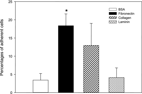
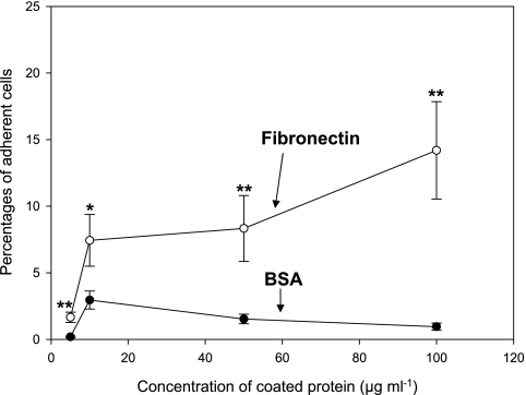
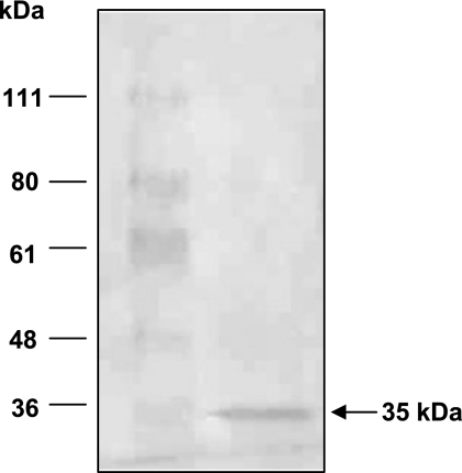
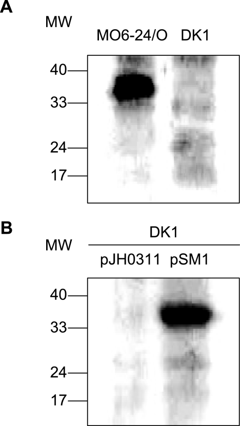
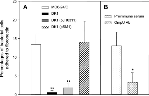
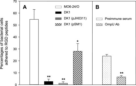
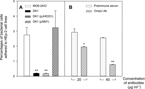
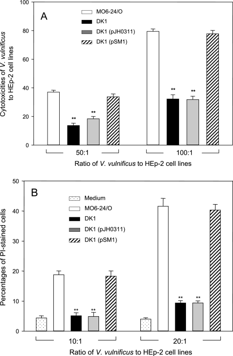
Vibrio vulnificus is a pathogenic bacterium that causes gastroenteritis and primary septicemia. To identify factors involved in microbial adherence to the host cells, we investigated bacterial proteins capable of binding to fibronectin, one of the main components comprised of the extracellular matrix of mammalian cells. A protein of approximately 35 kDa was purified from the extracts of V. vulnificus by its property to bind to immobilized fibronectin. This protein was identified as OmpU, one of the major outer membrane proteins of V. vulnificus. In binding assays using immobilized fibronectin, the number of ompU mutant cells bound to fibronectin was only 4% of that of wild-type cells bound to fibronectin. In addition, the exogenous addition of antibodies against OmpU resulted in a decreased ability of wild-type V. vulnificus to adhere to fibronectin. The ompU mutant was also defective in its adherence to RGD tripeptide (5% of the adherence of the wild type to RGD), cytoadherence to HEp-2 cells (7% of the adherence of the wild type to HEp-2), cytotoxicity to cell cultures (39% of the cytotoxicity of the wild type), and mortality in mice (10-fold increase in the 50% lethal dose). The ompU mutant complemented with the intact ompU gene restored its abilities for adherence to fibronectin, RGD tripeptide, and HEp-2 cells; cytotoxicity to HEp-2 cells; and mouse lethality. This study indicates that OmpU is an important virulence factor involved in the adherence of V. vulnificus to the host cells.


Figures

FIG. 1.
FIG. 1.
Adherence of Vibrio vulnificus to various components of the extracellular matrix. Wild-type V. vulnificus cells (1 × 10⁵) were added to a multiwell plate coated with one of three extracellular matrix proteins at a concentration of 100 μg ml−1 and incubated for 10 min. Assay of binding of V. vulnificus to each component was performed eight times. The percentages of bacterial cells that adhered to wells coated with BSA (an open bar), fibronectin (a black bar), collagen (a hatched bar), or laminin (a crosschecked bar) were determined by plating the eluted bacteria onto LBS agar. Error bars represent the means ± standard deviations from three independent experiments. The asterisk indicates a binding level that was significantly different from that of the BSA-coated control by Student's t test.
FIG. 2.
FIG. 2.
Adherence of Vibrio vulnificus to fibronectin- or BSA-coated surfaces. Wild-type V. vulnificus cells were added to the multiwell plate coated with various concentrations (5, 10, 50, and 100 μg ml−1) of fibronectin or BSA and incubated for 10 min. The percentages of bacterial cells that adhered to wells coated with BSA (filled circles) or fibronectin (open circles) were determined by counting CFU on LBS agar plates. Assay of binding of V. vulnificus to fibronectin or BSA at each concentration was performed eight times. Error bars represent the means ± standard deviations from three independent experiments. Asterisks indicate binding levels that were significantly different from those of the BSA-coated control at the corresponding concentration by the Student's t test. Data with P values of <0.01 are indicated with two asterisks, whereas data with P values of between 0.01 and 0.05 are indicated with one asterisk.
FIG. 3.
FIG. 3.
Identification of a fibronectin-binding protein of Vibrio vulnificus. A crude lysate of wild-type V. vulnificus was added to a multiwell plate coated with 50 μg of fibronectin and incubated for 3 h. After being washed with PBS, the proteins bound to the fibronectin-coated surface were eluted with 1% sodium dodecyl sulfate and concentrated fivefold with Centricon as described in Materials and Methods. Upon separation by 10% SDS-PAGE and transfer to a PVDF membrane, the eluted proteins were visualized by Ponceau. A protein of 35 kDa was excised and subjected to N-terminal amino acid sequencing.
FIG. 4.
FIG. 4.
Western blot analysis using polyclonal antibodies against the recombinant OmpU protein (anti-OmpU). (A) Confirmation of ompU mutant V. vulnificus by Western blot analysis. Lane 1, lysate of wild-type V. vulnificus MO6-24/O; lane 2, lysate of DK1, an ompU mutant of V. vulnificus. (B) Confirmation of the ompU complementation strain by Western blot analysis. Lane 1, lysate of DK1 harboring a broad-host-range vector, pJH0311; lane 2, lysate of DK1 harboring pSM1, a complementation plasmid of the wild-type ompU gene. Lysates of four different strains of V. vulnificus were prepared as described in Materials and Methods. Fifty micrograms of protein was subjected to SDS-PAGE per each sample and transferred onto a membrane. After being blocked with 5% nonfat dry milk, the membrane was incubated with mouse antibodies against the recombinant OmpU and was then incubated with horseradish peroxidase-conjugated anti-mouse immunoglobulin G. Immunoreactive proteins were visualized using an enhanced chemiluminescence system. MW, molecular weight (in thousands).
FIG. 5.
FIG. 5.
Role of the OmpU protein in adherence of Vibrio vulnificus to immobilized fibronectin. (A) Adherence of various V. vulnificus strains to fibronectin. V. vulnificus cells (wild-type V. vulnificus MO6-24/O [open bar], ompU mutant strain DK1 [closed bar], DK1 carrying pJH0311 [gray bar], and DK1 carrying a complementation plasmid, pSM1 [hatched bar]) were added to a 96-well plate coated with fibronectin. The percentage of bacterial cells that adhered to fibronectin-coated wells was determined by plating bound bacteria onto LBS agar. Error bars represent the means ± standard deviations from three independent experiments. Assay of the binding of each V. vulnificus strain to fibronectin was performed in quadruplicate for each experiment. Asterisks indicate binding levels that were significantly different from that of wild-type V. vulnificus by the Student's t test. Data with P values of <0.01 are indicated with two asterisks, whereas data with P values of between 0.01 and 0.05 are indicated with one asterisk. (B) Inhibition of fibronectin binding of wild-type V. vulnificus by anti-OmpU. The percentage of bacterial cells that adhered to fibronectin-coated wells was determined for wild-type V. vulnificus preincubated with mouse preimmune serum (dotted bar) or with anti-OmpU (cross-hatched bar) at 20 μg ml−1. Error bars represent the means ± standard deviations from three independent experiments. Assay of binding of V. vulnificus to fibronectin was performed in quadruplicate for each experiment. The asterisk indicates a binding level that was significantly different from that of wild-type V. vulnificus pretreated with mouse preimmune serum by Student's t test (0.01 < P < 0.05). Ab, antibody.
FIG. 6.
FIG. 6.
Role of OmpU protein in adherence of Vibrio vulnificus to immobilized RGD tripeptide. (A) Adherence of various V. vulnificus strains to the RGD tripeptide. V. vulnificus cells (wild-type V. vulnificus MO6-24/O [open bar], ompU mutant DK1 [closed bar], DK1 carrying pJH0311 [gray bar], and DK1 carrying a complementation plasmid, pSM1 [hatched bar]) were added to the 96-well plate coated with RGD tripeptide. Each well of the culture plate was coated with 20 μg of RGD tripeptide in PBS at 4°C for 18 h. V. vulnificus cells (1 × 10⁵) were added to each of the RGD-coated wells and incubated at RT for 30 min. The percentage of bacterial cells that adhered to RGD-coated wells was determined by counting CFU of the eluted bacteria on LBS agar plates. Error bars represent the means ± standard deviations from three independent experiments. Assay of binding of V. vulnificus to RGD tripeptide was performed in quadruplicate for each experiment. Asterisks indicate binding levels that were significantly different from that of wild-type V. vulnificus by Student's t test. Data with P values of <0.01 are indicated with two asterisks, whereas data with P values of between 0.01 and 0.05 are indicated with one asterisk. (B) Inhibition of RGD binding of wild-type V. vulnificus by anti-OmpU. The percentage of bacterial cells that adhered to RGD-coated wells was determined for wild-type V. vulnificus preincubated with mouse preimmune serum (dotted bar) or with anti-OmpU (cross-hatched bar) at 20 μg ml−1. Error bars represent the means ± standard deviations from three independent experiments. Assay of binding of V. vulnificus to RGD tripeptide was performed in quadruplicate for each experiment. Asterisks indicate binding levels that were significantly different (P < 0.01) from that of wild-type V. vulnificus pretreated with mouse preimmune serum by Student's t test. Ab, antibody.
FIG. 7.
FIG. 7.
Role of OmpU protein in adherence of Vibrio vulnificus to HEp-2 cells. (A) Cytoadherence of various V. vulnificus strains to HEp-2 cells. V. vulnificus cells (wild-type V. vulnificus MO6-24/O [open bar], ompU mutant DK1 [closed bar], DK1 carrying pJH0311 [gray bar], and DK1 carrying a complementation plasmid, pSM1 [hatched bar]) were added to HEp-2 cells at an MOI of 10 and incubated for 30 min. Following five washes with PBS, the HEp-2 cells were treated with 0.1% Triton X-100 for 15 min. The percentage of bacterial cells that adhered to HEp-2 was determined by measuring CFU of the retrieved bacteria on LBS agar plates. Assay of adherence of various V. vulnificus cells to HEp-2 was performed in quadruplicate, and data are presented with error bars, which are standard deviations of three independent experiments. Asterisks indicate binding levels that were significantly different from that of wild-type V. vulnificus by Student's t test. Data with P values of <0.01 are indicated with two asterisks, whereas data with P values of between 0.01 and 0.05 are indicated with one asterisk. (B) Inhibition of cytoadherence of wild-type V. vulnificus cells to HEp-2 cells by anti-OmpU. Prior to the cytoadherence test, wild-type V. vulnificus cells were treated with either antibodies against recombinant OmpU (OmpU Ab) (cross-hatched bars) or mouse preimmune serum (dotted bars) for 30 min at two different concentrations (20 or 40 μg ml−1). The percentage of bacterial cells that adhered to HEp-2 cells was determined by measuring CFU of the adhered bacteria on LBS plates. Each cytoadherence assay was done in quadruplicate, and data are presented along with standard deviations of three independent experiments. Asterisks indicate binding levels that were significantly different (P < 0.01) from that of wild-type V. vulnificus pretreated with mouse preimmune serum by the Student's t test.
FIG. 8.
FIG. 8.
Role of the OmpU protein of Vibrio vulnificus in bacterial cytotoxicity to HEp-2 cells. (A) Determination of cytotoxicity of various V. vulnificus strains by estimating the activity of LDH. Using a CytoTox96 assay kit, LDH released from the HEp-2 cells was measured upon incubation with wild-type V. vulnificus (open bars), ompU mutant DK1 (black bars), DK1 carrying pJH0311 (gray bars), or DK1 carrying pSM1 (hatched bars). HEp-2 cells (1 × 10⁵) were incubated with V. vulnificus cells for 30 min at two different MOIs, 50 and 100. Each assay was performed in quadruplicate and repeated three times. The data are shown with error bars, which are standard deviations of three independent experiments. Asterisks indicate enzyme activities that were significantly different (P < 0.01) from that of wild-type V. vulnificus by Student's t test. (B) Determination of the cytotoxicities of various V. vulnificus strains by staining with PI. The percentages of dead HEp-2 cells during incubation with wild-type V. vulnificus (open bars), ompU mutant strain DK1 (black bars), DK1 carrying pJH0311 (gray bars), or DK1 carrying pSM1 (hatched bars) were measured at two different MOIs, 10 and 20. HEp-2 cells not exposed to V. vulnificus were also stained with PI (dotted bars). Error bars represent the means ± standard deviations from three independent experiments. Asterisks indicate percentages of PI-stained cells that were significantly different (P < 0.01) from that of wild-type V. vulnificus cells by Student's t test.

References

    1. Aeckersberg, F., C. Lupp, B. Feliciano, and E. G. Ruby. 2001. Vibrio fischeri outer membrane protein OmpU plays a role in normal symbiotic colonization. J. Bacteriol. 183:6590-6597. - PMC - PubMed
    1. Bahrani, K., and J. D. Oliver. 1990. Studies on the lipopolysaccharide of virulent and avirulent strains of Vibrio vulnificus. Biochem. Cell Biol. 68:547-551. - PubMed
    1. Connell, T. D., A. J. Martone, and R. K. Holmes. 1995. A new mobilizable cosmid vector for use in Vibrio cholerae and other gram-negative bacteria. Gene 153:85-87. - PubMed
    1. Crawford, J. A., J. B. Kaper, and V. J. DiRita. 1998. Analysis of ToxR-dependent transcription activation of ompU, the gene encoding a major envelope protein in Vibrio cholerae. Mol. Microbiol. 29:235-246. - PubMed
    1. Di Martino, P. 2001. Effects of antibiotics on adherence of Pseudomonas aeruginosa and Pseudomonas fluorescens to human fibronectin. Chemotherapy 47:344-349. - PubMed

Publication types

LinkOut - more resources