. 2007 Feb;14(3):275-80.
doi: 10.1038/sj.gt.3302867. Epub 2006 Sep 21.

Optimization of cutaneous electrically mediated plasmid DNA delivery using novel electrode

Affiliations

Optimization of cutaneous electrically mediated plasmid DNA delivery using novel electrode

L C Heller et al. Gene Ther. 2007 Feb.

Abstract

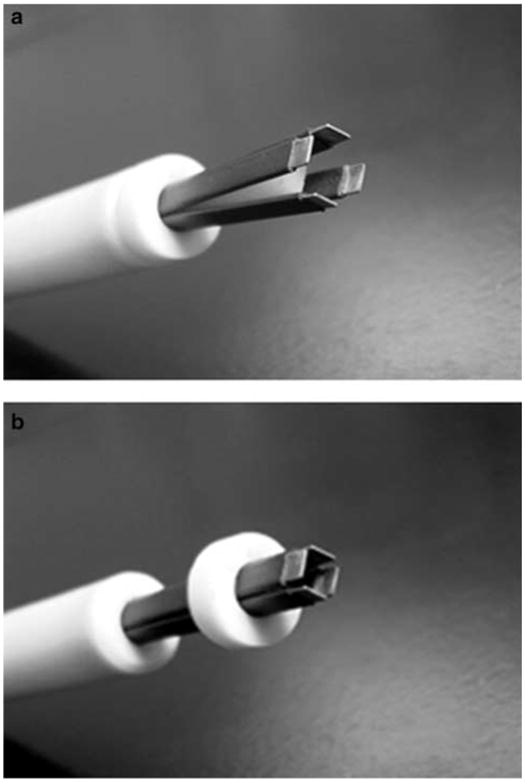
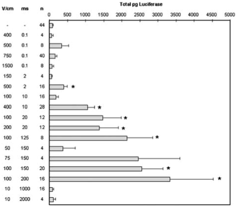
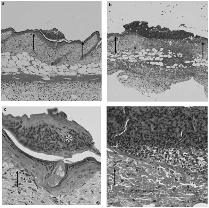
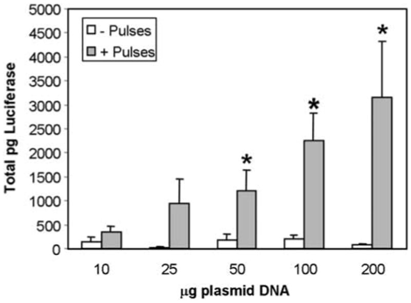
The easy accessibility of skin makes it an excellent target for gene transfer protocols. To take advantage of skin as a target for gene transfer, it is important to establish an efficient and reproducible delivery system. Electroporation is an established technique for enhancing plasmid delivery to many tissues in vivo. A critical component of this technique is the electrode configuration. Electroporation parameters were optimized for transgene expression with minimal tissue damage with a novel electrode. The highest transgene expression and efficiency of individual cell transformation with minimal damage was produced with eight 150 ms pulses at field strength of 100 V/cm. This electrode design offers the potential for easier and more reproducible electrically mediated cutaneous plasmid delivery than the simple electrodes currently commercially available. This electrode can be a valuable tool in determining the applicability of electrically mediated cutaneous gene transfer.


Figures

Figure 1
Figure 1
4PE: (a) open position; (b) closed position. A 6 mm gap is maintained between plates.
Figure 2
Figure 2
Comparison of luciferase expression after plasmid delivery with two types of pulsing conditions. The flank skin of 6–7 weeks old female C57/Bl6 mice (NCI) was shaved and then injected intradermally with 50 μl of 2 μg/μl pCMVLuc+ (a gift of Claude Nicolau, Harvard Medical School, Boston, MA, USA), which was prepared using Endo-free Megaprep kits (Qiagen, Valencia, CA, USA). Eight electric pulses were administered with a 90° rotation between sets of four pulses at a frequency of 1 Hz with a BTX T830 (BTX Molecular Delivery Systems, Holliston, MA, USA) using the 4PE electrode. Mice were anesthetized in an induction chamber charged with 3% isoflurane in O2 then fitted with a standard rodent mask and kept under general anesthesia during treatment. At 48 h after plasmid delivery, animals were humanely euthanized, and the tissue samples were excised and analyzed. For luciferase quantification, the tissue samples were homogenized in buffer (50 mM K3PO4, 1 mM EDTA, 1 mM DTT, 10% glycerol) using a Tissumizer (Tekmar, Cincinnati, OH, USA). Extracts were assayed for luciferase activity and quantified using an MLX microtiter plate luminometer (Dynex Technologies, Chantilly, VA, USA). Activity is expressed in total pg luciferase per tissue sample. Values represent mean and standard error. Due to the simplex scheme, some pulsing conditions were repeated multiple times as indicated. *P<0.05.
Figure 3
Figure 3
Combination of electroporation and electrophoresis. One hundred micrograms of pCMVLuc+ were delivered and luciferase assays were performed as described in Figure 2. Bars represent the mean of the means and standard error of the means for two replicate experiments. Each experiment contained four samples for each group. *P < 0.05 with respect to G+E-; **P < 0.05 with respect to electroporation pulse; ***P < 0.05 with respect to electrophoretic pulse.
Figure 4
Figure 4
Tissue damage following electroporation. Photographs of representative sections from study described in Table 2 illustrate examples of surface damage and subepidermal necrosis. (a and c) An area with surface damage is shown. (a) × 100 magnification and (c) × 400 magnification. (b and d) An area containing subepidermal necrosis is shown. (b) × 100 magnification and (d) × 400 magnification. Arrows point to the areas of normal tissue and stars are placed in the areas of damage.
Figure 5
Figure 5
Plasmid dose response. pCMVLuc+ (P) was injected intradermally in 50 μl at increasing concentrations, pulses (E) applied, and luciferase assays performed as described in Figure 2. Data represent mean and standard error of three separate experiments each containing four samples for each time point (total of 12 samples/time point). *P < 0.05

References

    1. Titomirov AV, Sukharev S, Kistanova E. In vivo electroporation and stable transformation of skin cells of newborn mice by plasmid DNA. Biochim Biophys Acta. 1991;1088:131–134. - PubMed
    1. Drabick JJ, Glasspool-Malone J, Somiari S, King A, Malone RW. Cutaneous transfection and immune responses to intradermal nucleic acid vaccination are significantly enhanced by in vivo electroporation. Mol Ther. 2000;3:249–255. - PubMed
    1. Glasspool-Malone J, Somiara S, Drabick JJ, Malone RW. Efficient nonviral cutaneous transfection. Mol Ther. 2000;2:140–146. - PubMed
    1. Chesnoy S, Huang L. Enhanced cutaneous gene delivery following intradermal injection of naked DNA in a high ionic strength solution. Mol Ther. 2001;5:57–62. - PubMed
    1. Maruyama H, Ataka K, Higuchi N, Sakamoto F, Gejyo F, Miyazaki J. Skin-targeted gene transfer using in vivo electroporation. Gene Therapy. 2001;8:1808–1812. - PubMed

Publication types