Comparative Study
. 2006 Oct 16;580(24):5612-20.
doi: 10.1016/j.febslet.2006.08.085. Epub 2006 Sep 12.

Quantitative comparison of the efficiency of antibodies against S1 and S2 subunit of SARS coronavirus spike protein in virus neutralization and blocking of receptor binding: implications for the functional roles of S2 subunit

Affiliations
Comparative Study

Quantitative comparison of the efficiency of antibodies against S1 and S2 subunit of SARS coronavirus spike protein in virus neutralization and blocking of receptor binding: implications for the functional roles of S2 subunit

Fanya Zeng et al. FEBS Lett. .

Abstract

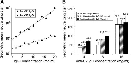
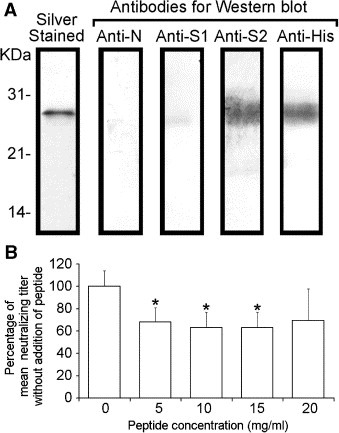
Neutralizing effects of antibodies targeting the C-terminal stalk (S2) subunit of the spike protein of severe acute respiratory syndrome coronavirus have previously been reported, although its mechanism remained elusive. In this study, high titered mouse antisera against the N-terminal globular (S1) and S2 subunits of the S protein were generated and total immunoglobulin G (IgG) was purified from these antisera. The efficiency of these purified IgGs in virus neutralization and blocking of receptor binding were compared quantitatively using virus neutralization assay and a previously developed cell-based receptor binding assay, respectively. We demonstrated that anti-S1 IgG neutralizes the virus and binds to the membrane associated S protein more efficiently than anti-S2 IgG does. Moreover, both anti-S1 and anti-S2 IgGs were able to abolish the binding between S protein and its cellular receptor(s), although anti-S1 IgG showed a significantly higher blocking efficiency. The unexpected blocking ability of anti-S2 IgG towards the receptor binding implied a possible role of the S2 subunit in virus docking process and argues against the current hypothesis of viral entry. On the other hand, the functional roles of the previously reported neutralizing epitopes within S2 subunit were investigated using an antigen specific antibody depletion assay. Depletion of antibodies against these regions significantly diminished, though not completely abolished, the neutralizing effects of anti-S2 IgG. It suggests the absence of a major neutralizing domain on S2 protein. The possible ways of anti-S2 IgGs to abolish the receptor binding and the factors restricting anti-S2 IgGs to neutralize the virus are discussed.


Conflict of interest statement

No conflicts declared.

Figures

Figure 1
Figure 1
Schematic diagram of S2 functional domains and regions containing neutralizing epitopes. The black boxes, white boxes and black lines represent the S2 functional domains, regions chosen for this study (designated as S2‐Ept) and regions containing previously reported neutralizing epitopes, respectively. The number of the references of the reported neutralizing regions is listed on the right column. FP, N‐HR, C‐HR and T refer to fusion peptide, N‐terminal and C‐terminal heptad repeat, and transmembrane domain respectively.
Figure 2
Figure 2
The neutralizing efficiency and additive neutralizing effect of anti‐S1 and anti‐S2 IgG. (A) The geometric mean neutralizing titers of anti‐S1 and anti‐S2 IgGs of the same concentration range, i.e. 2–20 mg/ml, were compared. The values of y‐axis are showed in logarithmic scale and the dotted lines represent the best‐fit line obtained from linear regression of the datasets. Data of Anti‐N IgG is not shown since it did not show any neutralizing effect in all tested concentrations. (B) The geometric mean neutralizing titers of anti‐S2 IgG of different concentrations mixed with 0.5 or 2 mg/ml of anti‐S1 IgG. The average values of the triplicates were indicated and the error bars represent the SD of the triplicates. The geometric mean neutralizing titers of 0.5 and 2 mg/ml of anti‐S1 IgG are 29.6 (S.D. = 3.8) and 51.3 (S.D. = 11.8), respectively.
Figure 3
Figure 3
The binding efficiency of anti‐S1 and anti‐S2 IgGs towards m‐S protein. (A) Flow cytometry showing that anti‐S1 and anti‐S2 IgGs specifically bind to m‐S protein on AD293 cells. The shaded peaks represent the background signal, i.e. cells without IgG incubation, and the non‐shaded peaks represent cells incubated with 0.8 (upper panel) or 3.2 (lower panel) mg/ml of anti‐N, anti‐S1 and anti‐S2 IgG as indicated. (B) Anti‐S1 IgG binds more efficiently to m‐S protein than anti‐S2 IgG does. The error bars represent the SD of the triplicates. (C) The results of the serial antibody binding and depleting assay. The upper and lower panels show the decrement of MFI and percentage of remaining IgG after each round of depletion, respectively. Each data point represents the average value of three triplicates.
Figure 4
Figure 4
Abolition of receptor binding by anti‐S1 and anti‐S2 IgGs in cell‐based receptor binding assay. (A) The abolition of receptor binding by anti‐S1 and anti‐S2 IgGs is specific. Ligand cells, i.e. AD293‐S/GFP, incubated with anti‐S1 and anti‐S2 IgGs showed a substantially smaller number of bound cells (green fluorescent spots) when compared with those incubated with anti‐N IgG or no IgG incubation. The images shown are representative views (magnification of 100×) of each treatment as indicated. (B) Abolition of receptor binding of different types of ligand cells by anti‐S2 IgGs. The significance of differences between the numbers of bound cells of the control IgG, i.e. anti‐N IgG, and anti‐S1 or anti‐S2 IgG is represented by asterisks (P < 0.01, ∗∗ or P < 0.05, ∗). The results are presented in average of the triplicates with S.D. The types of ligand cells used are indicated at the top of each data group. (C) The blocking efficiency of anti‐S1 IgG is higher than that of anti‐S2 IgG under the same concentrations tested. Each data point represents the average value of three replicates. The x‐axis, i.e. IgG concentration, is presented in logarithmic scale.
Figure 5
Figure 5
The role of S2‐Ept in virus neutralization. (A) Identification of S2‐Ept peptide using Western blot. (B) The neutralizing titer of S2‐Ept depleted anti‐S2 IgG is lower than the mock depleted anti‐S2 IgG (0 mg/ml). The results were presented as the average of geometric mean of neutralizing titer of three replicates and the error bar represents its SD. The statistical significance of differences between the geometric mean of neutralizing titer of dep‐anti‐S2 IgG and mock depleted anti‐S2 IgG is represented by an asterisk (P < 0.05, ∗). It is noted that no significant decrease of neutralizing titer was observed between dep‐anti‐S1 IgG and mock depleted anti‐S1 IgG (data not shown).

Similar articles

Cited by

References

    1. Peiris J.S., Lai S.T., Poon L.L., Guan Y., Yam L.Y., Lim W., Nicholls J., Yee W.K., Yan W.W., Cheung M.T., Cheng V.C., Chan K.H., Tsang D.N., Yung R.W., Ng T.K., Yuen K.Y., Coronavirus as a possible cause of severe acute respiratory syndrome. Lancet, 361, (2003), 1319– 1325. - PMC - PubMed
    1. Cavanagh D., Severe acute respiratory syndrome vaccine development: experiences of vaccination against avian infectious bronchitis coronavirus. Avian Pathol., 32, (2003), 567– 582. - PMC - PubMed
    1. Saif L.J., Animal coronavirus vaccines: lessons for SARS. Dev. Biol. (Basel), 119, (2004), 129– 140. - PubMed
    1. Taylor D.R., Obstacles and advances in SARS vaccine development. Vaccine, 24, (2006), 863– 871. - PMC - PubMed
    1. He Y., Jiang S., Vaccine design for severe acute respiratory syndrome coronavirus. Viral Immunol., 18, (2005), 327– 332. - PubMed

Publication types

MeSH terms