

The different cleavage DNA sequence specificity explains the camptothecin resistance of the human topoisomerase I Glu418Lys mutant
- PMID: 16990249
- PMCID: PMC1636438
- DOI: 10.1093/nar/gkl670
The different cleavage DNA sequence specificity explains the camptothecin resistance of the human topoisomerase I Glu418Lys mutant
Abstract
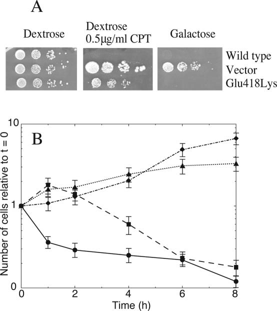
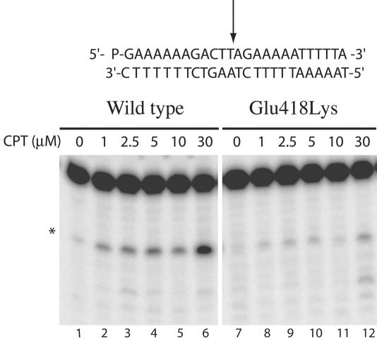
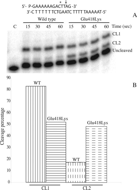
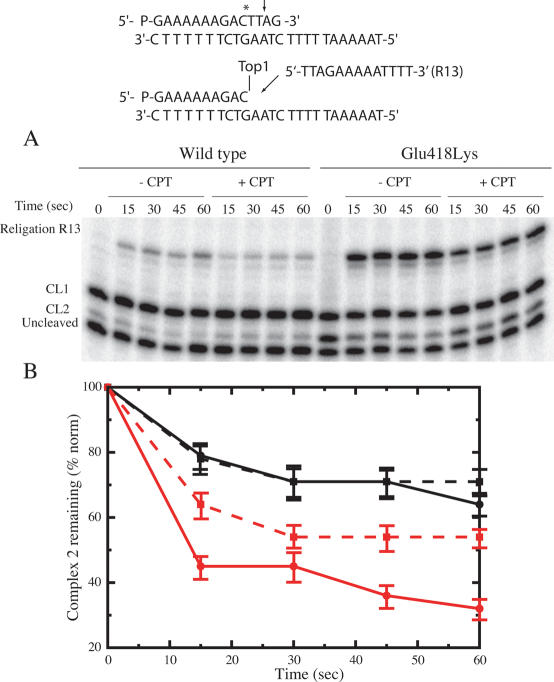
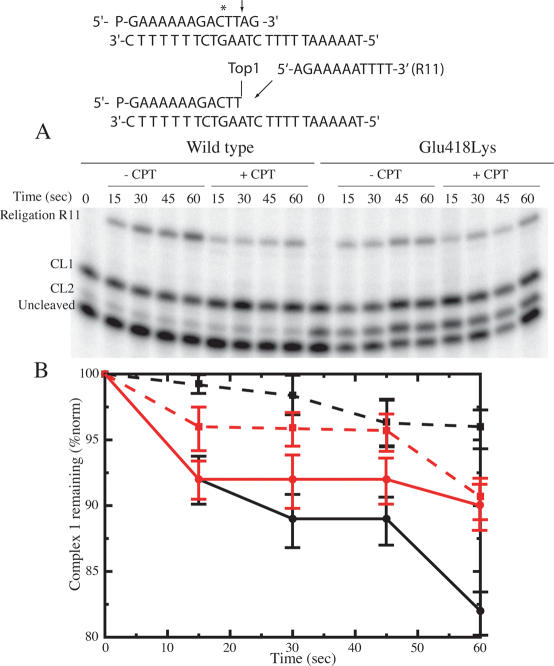
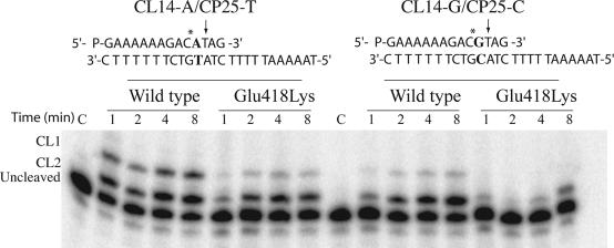
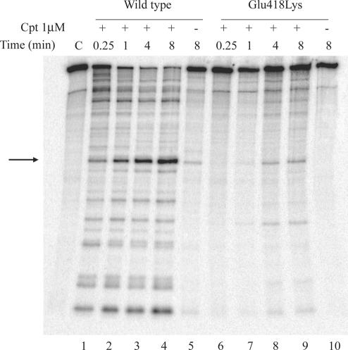
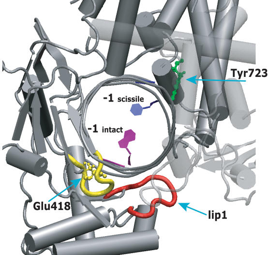
Yeast cells expressing the Glu418Lys human topoisomerase I mutant display a camptothecin resistance that slowly decreases as a function of time. Molecular characterization of the single steps of the catalytic cycle of the purified mutant indicates that it has a relaxation activity identical to the wild-type protein but a different DNA sequence specificity for the cleavage sites when compared to the wild-type enzyme, as assayed on several substrates. In particular the mutant has a low specificity for CPT sensitive cleavable sites. In fact, the mutant has, at variance of the wild-type enzyme, a reduced preference for cleavage sites having a thymine base in position -1 of the scissile strand. This preference, together with the strict requirement for a thymine base in position -1 for an efficient camptothecin binding, explains the temporary camptothecin resistance of the yeast cell expressing the mutant and points out the importance of the DNA sequence in the binding of the camptothecin drug.
Figures
Similar articles
-
Differential induction of topoisomerase I-DNA cleavage complexes by the indenoisoquinoline MJ-III-65 (NSC 706744) and camptothecin: base sequence analysis and activity against camptothecin-resistant topoisomerases I.Cancer Res. 2003 Nov 1;63(21):7428-35. Cancer Res. 2003. PMID: 14612542
-
The camptothecin-resistant topoisomerase I mutant F361S is cross-resistant to antitumor rebeccamycin derivatives. A model for topoisomerase I inhibition by indolocarbazoles.Biochemistry. 1999 Jul 6;38(27):8605-11. doi: 10.1021/bi983052y. Biochemistry. 1999. PMID: 10393535
-
Camptothecin resistance from a single mutation changing glycine 363 of human DNA topoisomerase I to cysteine.Cancer Res. 1993 Sep 15;53(18):4343-8. Cancer Res. 1993. PMID: 8395982
-
Using yeast to understand drugs that target topoisomerases.Ann N Y Acad Sci. 1996 Dec 13;803:32-43. doi: 10.1111/j.1749-6632.1996.tb26374.x. Ann N Y Acad Sci. 1996. PMID: 8993498 Review. No abstract available.
-
Domains of human topoisomerase I and associated functions.Prog Nucleic Acid Res Mol Biol. 1998;60:111-32. doi: 10.1016/s0079-6603(08)60891-0. Prog Nucleic Acid Res Mol Biol. 1998. PMID: 9594573 Review.
Cited by
-
Parallel Genomic Alterations of Antigen and Payload Targets Mediate Polyclonal Acquired Clinical Resistance to Sacituzumab Govitecan in Triple-Negative Breast Cancer.Cancer Discov. 2021 Oct;11(10):2436-2445. doi: 10.1158/2159-8290.CD-21-0702. Epub 2021 Aug 17. Cancer Discov. 2021. PMID: 34404686 Free PMC article.
-
Natural Compounds as Therapeutic Agents: The Case of Human Topoisomerase IB.Int J Mol Sci. 2021 Apr 16;22(8):4138. doi: 10.3390/ijms22084138. Int J Mol Sci. 2021. PMID: 33923641 Free PMC article. Review.
-
Swapping of The N-Terminal Domain of Human Topoisomerase 1B with the Corresponding Plasmodium Falciparum Counterpart Strongly Impairs Enzyme Activity.Rep Biochem Mol Biol. 2020 Jan;8(4):366-375. Rep Biochem Mol Biol. 2020. PMID: 32582794 Free PMC article.
-
Topoisomerase I gene mutations at F270 in the large subunit and N184 in the small subunit contribute to the resistance mechanism of the unicellular parasite Leishmania donovani towards 3,3'-diindolylmethane.Antimicrob Agents Chemother. 2009 Jun;53(6):2589-98. doi: 10.1128/AAC.01648-08. Epub 2009 Mar 30. Antimicrob Agents Chemother. 2009. PMID: 19332675 Free PMC article.
-
Topoisomerase IB: a relaxing enzyme for stressed DNA.Cancer Drug Resist. 2020 Mar 19;3(1):18-25. doi: 10.20517/cdr.2019.106. eCollection 2020. Cancer Drug Resist. 2020. PMID: 35582040 Free PMC article. Review.
References
-
- Chen A., Liu L.F. DNA topoisomerase: essential enzymes and lethal targets. Ann. Rev. Pharmacol. Toxicol. 1994;34:191–218. - PubMed
-
- Nitiss J. Investigating the biological functions of DNA topoisomerases in eukaryotic cells. Biochim. Biophys. Acta. 1998;1400:63–82. - PubMed
-
- Wang J.C. DNA topoisomerases. Ann. Rev. Biochem. 1996;65:635–692. - PubMed
-
- Stewart L., Ireton G.C., Champoux J.J. The domain organization of human topoisomerase I. J. Biol. Chem. 1996;271:7602–7608. - PubMed
-
- Redinbo M.R., Stewart L., Kuhn P., Champoux J.J., Hol W.G.J. Crystal structures of human topoisomerase I in covalent and noncovalent complexes with DNA. Science. 1998;279:1504–1513. - PubMed
