

A Trojan horse transition state analogue generated by MgF3- formation in an enzyme active site
- PMID: 16990434
- PMCID: PMC1595420
- DOI: 10.1073/pnas.0604448103
A Trojan horse transition state analogue generated by MgF3- formation in an enzyme active site
Abstract
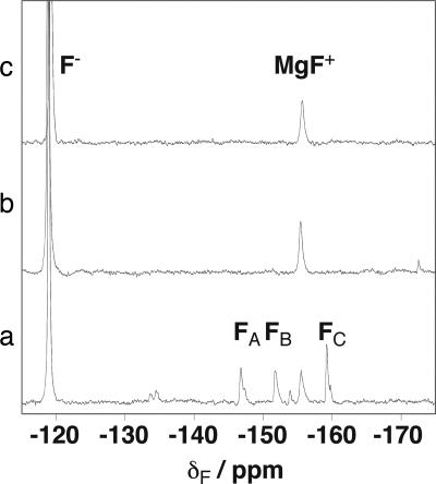
Identifying how enzymes stabilize high-energy species along the reaction pathway is central to explaining their enormous rate acceleration. beta-Phosphoglucomutase catalyses the isomerization of beta-glucose-1-phosphate to beta-glucose-6-phosphate and appeared to be unique in its ability to stabilize a high-energy pentacoordinate phosphorane intermediate sufficiently to be directly observable in the enzyme active site. Using (19)F-NMR and kinetic analysis, we report that the complex that forms is not the postulated high-energy reaction intermediate, but a deceptively similar transition state analogue in which MgF(3)(-) mimics the transferring PO(3)(-) moiety. Here we present a detailed characterization of the metal ion-fluoride complex bound to the enzyme active site in solution, which reveals the molecular mechanism for fluoride inhibition of beta-phosphoglucomutase. This NMR methodology has a general application in identifying specific interactions between fluoride complexes and proteins and resolving structural assignments that are indistinguishable by x-ray crystallography.
Conflict of interest statement
The authors declare no conflict of interest.
Figures


Similar articles
-
Kinetic analysis of beta-phosphoglucomutase and its inhibition by magnesium fluoride.J Am Chem Soc. 2009 Feb 4;131(4):1575-88. doi: 10.1021/ja806421f. J Am Chem Soc. 2009. PMID: 19132841
-
MgF(3)(-) and alpha-galactose 1-phosphate in the active site of beta-phosphoglucomutase form a transition state analogue of phosphoryl transfer.J Am Chem Soc. 2009 Nov 18;131(45):16334-5. doi: 10.1021/ja905972m. J Am Chem Soc. 2009. PMID: 19852484
-
Catalytic cycling in beta-phosphoglucomutase: a kinetic and structural analysis.Biochemistry. 2005 Jul 12;44(27):9404-16. doi: 10.1021/bi050558p. Biochemistry. 2005. PMID: 15996095
-
Metal Fluorides: Tools for Structural and Computational Analysis of Phosphoryl Transfer Enzymes.Top Curr Chem (Cham). 2017 Apr;375(2):36. doi: 10.1007/s41061-017-0130-y. Epub 2017 Mar 15. Top Curr Chem (Cham). 2017. PMID: 28299727 Free PMC article. Review.
-
Phosphoryl transfer enzymes and hypervalent phosphorus chemistry.Acc Chem Res. 2004 Oct;37(10):746-53. doi: 10.1021/ar040053b. Acc Chem Res. 2004. PMID: 15491121 Review.
Cited by
-
1H, 15N and 13C backbone resonance assignments of the P146A variant of β-phosphoglucomutase from Lactococcus lactis in its substrate-free form.Biomol NMR Assign. 2019 Oct;13(2):349-356. doi: 10.1007/s12104-019-09904-y. Epub 2019 Aug 8. Biomol NMR Assign. 2019. PMID: 31396843 Free PMC article.
-
Insights into the reaction of protein-tyrosine phosphatase 1B: crystal structures for transition state analogs of both catalytic steps.J Biol Chem. 2010 May 21;285(21):15874-83. doi: 10.1074/jbc.M109.066951. Epub 2010 Mar 16. J Biol Chem. 2010. PMID: 20236928 Free PMC article.
-
Briefly bound to activate: transient binding of a second catalytic magnesium activates the structure and dynamics of CDK2 kinase for catalysis.Structure. 2011 May 11;19(5):675-90. doi: 10.1016/j.str.2011.02.016. Structure. 2011. PMID: 21565702 Free PMC article.
-
Peri active site catalysis of proline isomerisation is the molecular basis of allomorphy in β-phosphoglucomutase.Commun Biol. 2024 Jul 27;7(1):909. doi: 10.1038/s42003-024-06577-9. Commun Biol. 2024. PMID: 39068257 Free PMC article.
-
Atomic details of near-transition state conformers for enzyme phosphoryl transfer revealed by MgF-3 rather than by phosphoranes.Proc Natl Acad Sci U S A. 2010 Mar 9;107(10):4555-60. doi: 10.1073/pnas.0910333106. Epub 2010 Feb 17. Proc Natl Acad Sci U S A. 2010. PMID: 20164409 Free PMC article.
References
Publication types
MeSH terms
Substances
LinkOut - more resources
Full Text Sources
