

Amino acids 1 to 422 of the spike protein of SARS associated coronavirus are required for induction of cyclooxygenase-2
- PMID: 16991002
- PMCID: PMC7088811
- DOI: 10.1007/s11262-005-0070-4
Amino acids 1 to 422 of the spike protein of SARS associated coronavirus are required for induction of cyclooxygenase-2
Abstract
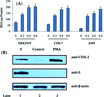
The causative agent of severe acute respiratory syndrome (SARS) has been identified as SARS-associated coronavirus (SARS-CoV). To evaluate the molecular mechanisms involved in the viral infection, in this study, we investigated the role of SARS-CoV Spike (S) protein in the regulation of cyclooxygenase-2 (COX-2). Expression of COX-2 stimulated by the S protein was verified by RT-PCR and western blot assay. To explore the relationship between S and COX-2, we constructed a series of plasmids containing truncated N-terminal fragments of the SARS-CoV S gene (designated from Sa to Si), which encoded truncated S proteins, and investigated whether these truncated proteins could induce effective expression of COX-2 in 293T cells. Our results showed that S(d) that encoded a truncated S protein with 422 amino acid residues (from 1 to 422 aa), a part of 672 amino-acid S1 subunit is crucial for the induction of COX-2 expression. Immunofluorescence examinations also give the evidence that these N terminal 422 amino acids of the S protein were also required for the correct localization of the protein. We also compared S protein sequences of SARS-CoV isolated during the SARS break with that from palm civets, a possible source of SARS-CoV found in humans. S protein residues (344, 360), which mutated in the epitome from palm civet to human being were characterized in 3D modeling of 252-375 amino acid fragment. Collectively, these results indicate that S protein of SARS-CoV induces the expression of COX-2 and an N-terminal fragment of the Spike protein is crucial for the induction. Our finding may provide clue for the induction of inflammation by SARS-CoV and cast insight into the severity of the SARS epidemic.
Figures
References
-
- Rota P.A., Oberste M.S., Monroe S.S., Nix W.A., Campagnoli R., Icenogle J.P., Penaranda S., Bankamp B., Maher K., Chen M.H., Tong S., Tamin A., Lowe L., Frace M., DeRisi J.L., Chen Q., Wang D., Erdman D.D., Peret T.C., Burns C., Ksiazek T.G., Rollin P.E., Sanchez A., Lick S., Holloway B., Limor J., McCaustland K., Olsen-Rasmussen M., Fouchier R., Gunther S., Osterhaus A.D., Drosten C., Pallansch M.A., Anderson L.J., Bellini W.J. Science. 2003;300(5624):1394–1399. doi: 10.1126/science.1085952. - DOI - PubMed
-
- Marra M.A., Jones S.J., Astell C.R., Holt R.A., Brooks-Wilson A., Buttereld Y.S., Khattra J., Asano J.K., Barber S.A., Chan S.Y., Cloutier A., Coughlin S.M., Freeman D., Girn N., Grith O.L., Leach S.R., Mayo M., McDonald H., Montgomery S.B., Pandoh P.K., Petrescu A.S., Robertson A.G., Schein J.E., Siddiqui A., Smailus D.E., Stott J.M., Yang G.S., Plummer F., Andonov A., Artsob H., Bastien N., Bernard K., Booth T.F., Bowness D., Czub M., Drebot M., Fernando L., Flick R., Garbutt M., Gray M., Grolla A., Jones S., Feldmann H., Meyers A., Kabani A., Li Y., Normand S., Stroher U., Tipples G.A., Tyler S., Vogrig R., Ward D., Watson B., Brunham R.C., Krajden M., Petric M., Skowronski D.M., Upton C., Roper R.L. Science. 2003;300:1399–1404. doi: 10.1126/science.1085953. - DOI - PubMed
Publication types
MeSH terms
Substances
LinkOut - more resources
Full Text Sources
Research Materials
Miscellaneous
