. 1990 Nov;5(5):569-81.
doi: 10.1016/0896-6273(90)90212-x.

A family of glutamate receptor genes: evidence for the formation of heteromultimeric receptors with distinct channel properties

Affiliations

A family of glutamate receptor genes: evidence for the formation of heteromultimeric receptors with distinct channel properties

N Nakanishi et al. Neuron. 1990 Nov.

Abstract

We have isolated two cDNA clones (GluR-K2 and GluR-K3) that share considerable sequence identity with the previously described glutamate receptor subunit, GluR-K1. The three glutamate receptor subunits show significant sequence conservation with the glutamine binding component of the glutamine permease of E. coli. Each of these clones encodes a channel responsive to both kainate and AMPA. The coexpression of GluR-K2 with either GluR-K3 or GluR-K1 results in the formation of channels whose current-voltage relationships differ from those of the individual subunits alone and more closely approximate the properties of kainate receptors in neurons. These observations indicate that the kainate/quisqualate receptors are encoded by a family of genes and are likely to be composed of hetero-oligomers of at least two distinct subunits.


Figures
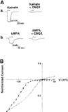
Figure 1
Figure 1. Expression of Functional Kainate/AMPA-Responsive Channels in Xenopus Oocytes after Injection of Synthetic GluR-K1 mRNA
(A) Trace a: Voltage-clamp recording from a single Xenopus oocyte injected with 3 ng of synthetic GluR-K1 RNA. Downward deflections indicate inward currents. The oocyte membrane potential was maintained at −100 mV, and kainate (100 µM) was applied by superfusion for the duration indicated by the bars. The antagonist CNQX (10 µM) reversibly blocked the GluR-K1 kainate response (recovery not shown). Trace b: Current traces obtained from the same egg after application of 10 µM AMPA for the duration indicated. The response was reversibly blocked by CNQX. (B) Current-voltage plot of the kainate (200 µM)-evoked response of oocytes injected with 3 ng of GluR-K1 (closed circles) or 50 ng of rat cerebral cortex and hippocampal poly(A)+ mRNA (open circles). Responses were normalized to current values evoked at a holding potential of −150 mV. Each point represents the mean and standard error of kainate-evoked currents obtained from 4–7 oocytes.
Figure 2
Figure 2. Alignment of the Deduced Ammo Acid Sequences of the Rat GluR-K2 and GluR-K3 cDNAs with Published Sequences of GluR-K1, Chick KBP, and Frog KBP
The predicted amino-terminal residue of the mature protein is numbered 1, and the preceding residues are indicated by negative numbers. Boxed amino acid residues are those identical in all three of the GluR-K subunits. The four proposed transmembrane domains (M1-M4) are indicated by solid bars. The two sets of arrows denote the borders of two regions of the GluR-K subunits and the KBPs that bear homology to E. coli GlnH (see Figure 3). Smaller residues conserved within the putative M2 region are shaded. Triangles indicate the position of negatively charged residues flanking the M2 regions of the GluR-K subunits. The deduced amino acid sequence for GluR-K1 is from Hollmann et al. (1989); that for chick KBP, from Gregor et al. (1989); and that for frog KBP, from Wada et al. (1989). The DNA sequences of the GluR-K2 and GluR-K3 cDNAs are available on request and have been deposited with the GenBank-EMBL database under accession numbers X54655 and X54656, respectively.
Figure 3
Figure 3. Sequence Homology between E. coli GlnH, GluR-K Subunits, and Two KBPs
(A) Sequence homology between E. coli GlnH (amino acid residues 4-98; Nohno et al., 1986), GluR-K subunits, and two KBPs within the region indicated by the first set of arrows in Figure 2. Exact positions in each protein are shown by numbers in parentheses. Boxed amino acid residues represent identity between GlnH and at least one of the GluR-K subunits or KBPs. In this region, sequence identity between GlnH and each of the kainate receptor subunits is 33% for GluR-K2, 30% for GluR-K3, 30% for GluR-K1, 21% for chick KBP, and 29% for frog KBK. The alignment shown in this figure gives a similarity score that is 12.7 standard deviations above the mean value of those obtained from the alignments of 200 random shuffles of the GluR-K1 sequence (amino acids 389-504) with GlnH. Two gaps were introduced to maximize identities. (B) Sequence homology between amino acid residues 176 and 220 of E. coli GlnH, GluR-K subunits, and two KBPs in the region indicated by the second set of arrows in Figure 2. In this region, sequence identity between GlnH and each of the kainate receptor subunits is 44% for GluR-K2, 42% for GluR-K3, 47% for GluR-K1, 29% for chick KBP, and 27% for frog KBP. The alignment shown in this figure gives a similarity score that is 12.0 standard deviations above the mean value of those obtained from the alignments of 200 random shuffles of the GluR-K1 sequence (amino acids 719-762) with GlnH.
Figure 4
Figure 4. GluR-K2 and GluR-K3 Generate Kainate- and AMPA-Sensitive Channels when Expressed Alone in Xenopus Oocytes
(A) Inward current responses are shown to kainate (100 µM) and AMPA (10 µM) 4–5 days after injection of 10 ng of GluR-K2 (trace a) or 3 ng of GluR-K3 (trace b) mRNA. Membrane potentials were held at −110 mV for GluR-K2. For GluR-K3, the egg was clamped at −70 mV (kainate) and −100 mV (AMPA). All kainate and AMPA responses were inhibited by the antagonist CNQX (10 µM). The same egg injected with either GluR-K2 or GluR-K3 mRNA was used to record the kainate- and AMPA-evoked currents shown in trace a (GluR-K2) and trace b (GluR-K3). (B) Dose-response curves of GluR-K3 for kainate (closed triangles) and AMPA (closed circles) compared with those of GluR-K1 for kainite (open triangles) and AMPA (open circles). Responses were normalized to average current values evoked at concentrations of 300 µM AMPA and 3 mM kainate. Each point represents the mean and standard error of agonist-evoked currents obtained from 3–6 oocytes, voltage-clamped at −70 mV. EC50 values and Hill coefficients derived from curves are listed in Table 1. Curves were fitted as described in Experimental Procedures. (C) Current-voltage plot of kainate (200 µM) currents evoked in oocytes injected with GluR-K3 RNA (3 ng) (open circles) compared with that obtained from oocytes injected with GluR-K1 RNA (3 ng) (closed circles). Responses were normalized to current values evoked at −150 mV. Each point represents the mean and standard error of kainate-evoked currents obtained from 4–6 oocytes.
Figure 5
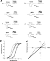
Figure 5. Combinations of GluR-K Subunits Form Distinct Channels when Expressed in Xenopus Oocytes
(A) Current traces recorded from single oocytes 4–5 days after injection with GluR-K1 + GluR-K3 (trace a), GluR-K1 + GluR-K2 (trace b), GluR-K3 + GluR-K2 (trace c), or GluR-K1 + GluR-K2 + GluR-K3 (trace d) mRNA. Oocytes were injected with 3 ng of RNA from each subunit (6 ng total for paired combinations, 9 ng for GluR-K1 + GluR-K2 + GluR-K3). Membrane potentials were held at −70 mV, except for the GluR-K1 + GluR-K2 AMKA (−100 mV) and GluR-K1 + CluR-K2 + GluR-K3 kainate (ȡ50 mV) responses. Kainate (100 µM) or AMPA (10 µM) was applied through the bath for the durations indicated by the bars. Responses were blocked by CNQX (10 µM). For all subunit combinations, the same egg was used to record the kainate- and AMPA-evoked responses shown. (B) Kainate dose-response curves for GluR-K1 + GluR-K3 (closed triangles), GluR-K1 + GluR-K2 (closed circles), GluR-K3 + GluR-K2 (closed diamonds), and GluR-K1 + GluR-K2 + GluR-K3 (closed squares) together with AMPA dose-response curves for GluR-K1 + GluR-K3 (open triangles), GluR-K1 + GluR-K2 (open circles), GluR-K3 + GluR-K2 (open diamonds), and GluR-K1 + GluR-K2 + GluR-K3 (open squares). For each curve, responses were normalized to average current values evoked at concentrations of 300 µM AMPA and 3 mM kainate. Each point represents the mean and standard error of agonist-evoked currents obtained from 4–6 oocytes and recorded at a holding potential of −70 mV. EC50 values and Hill coefficients derived from these curves are listed in Table 1. (C) Current-voltage plots of kainate currents evoked in oocytes injected with GluR-K1 + GluR-K3 (closed triangles), GluR-K1 + GluR-K2 (closed circles), GluR-K3 + GluR-K2 (open squares), or GluR-K1 + GluR-K2 + GluR-K3 (open circles). Oocytes were injected with 3 ng of each subunit RNA. Responses were normalized to current values evoked at −150 mV. Each point represents the mean and standard error of kainate-evoked currents obtained from 4–7 oocytes.

Similar articles

Cited by

References

    1. Ames GF-L. Bacterial periplasmic transport systems: structure, mechanism, and evolution. Annu. Rev. Biochem. 1986;55:397–425. - PubMed
    1. Ascher P, Nowak L. Quisqualate and kainate-activated channels in mouse central neurons in culture. J. Physiol. 1988;399:227–245. - PMC - PubMed
    1. Chomczynski P, Sacchi N. Single-step method of RNA isolation by acid guanidinium thiocyanate-phenol-chloroform extraction. Anal. Biochem. 1987;162:156–159. - PubMed
    1. Christie MI, North RA, Osborne PB, Douglass J, Adelman JP. Heteropolymeric potassium channels expressed in Xenopus oocytes from cloned subunits. Neuron. 1990;4:405–411. - PubMed
    1. Claudio T, Ballivet M, Patrick J, Heinemann S. Nucleotide and deduced amino acid sequence of Torpedo cafifornica acetylcholine receptor gamma subunit. Proc. Natl. Acad. Sci. USA. 1983;80:1111–1115. - PMC - PubMed

Publication types

MeSH terms

Associated data