. 2006 Nov 15;299(2):386-97.
doi: 10.1016/j.ydbio.2006.08.002. Epub 2006 Aug 9.

Interaction of PAR-6 with CDC-42 is required for maintenance but not establishment of PAR asymmetry in C. elegans

Affiliations

Interaction of PAR-6 with CDC-42 is required for maintenance but not establishment of PAR asymmetry in C. elegans

Donato Aceto et al. Dev Biol. .

Abstract

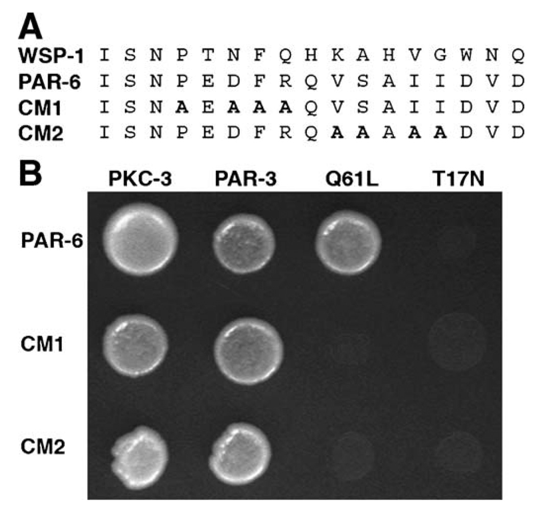
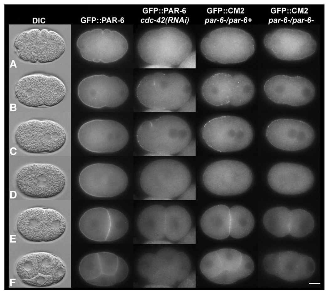
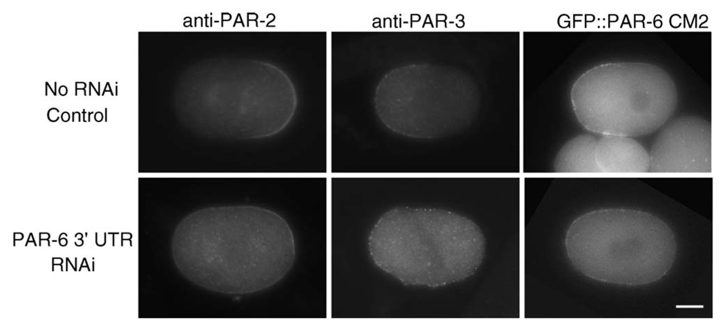
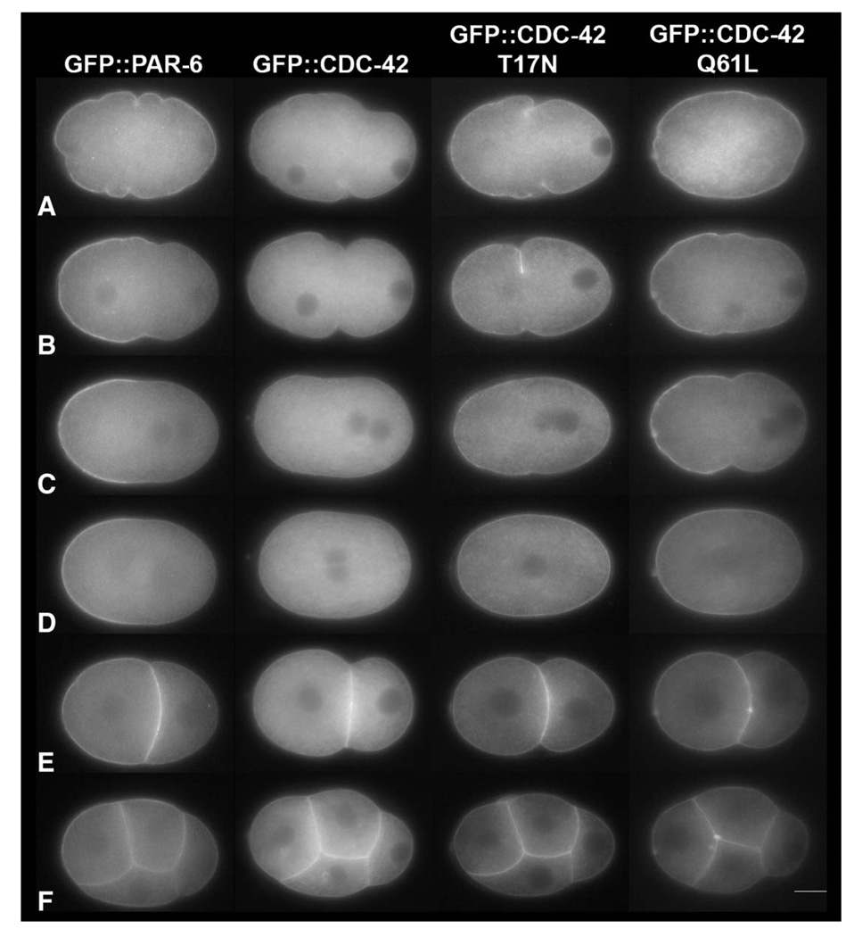
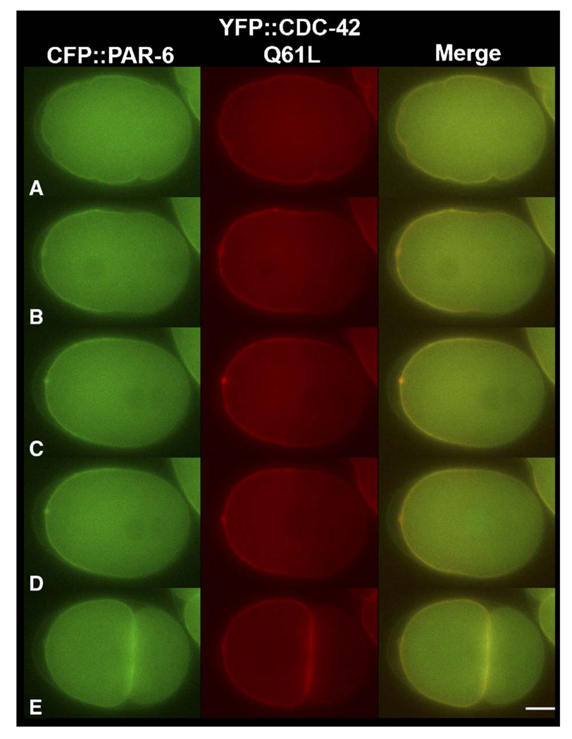
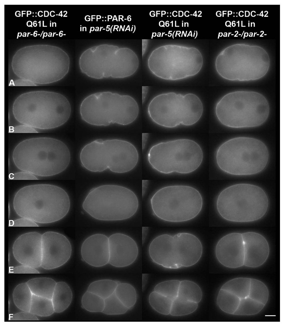
Caenorhabditis elegans embryonic polarity requires the asymmetrically distributed proteins PAR-3, PAR-6 and PKC-3. The rho family GTPase CDC-42 regulates the activities of these proteins in mammals, flies and worms. To clarify its mode of action in C. elegans we disrupted the interaction between PAR-6 and CDC-42 in vivo, and also determined the distribution of GFP-tagged CDC-42 in the early embryo. Mutant PAR-6 proteins unable to interact with CDC-42 accumulated asymmetrically, at a reduced level, but this asymmetry was not maintained during the first division. We also determined that constitutively active GFP::CDC-42 becomes enriched in the anterior during the first cell cycle in a domain that overlaps with PAR-6. The asymmetry is dependent on PAR-2, PAR-5 and PAR-6. Furthermore, we found that overexpression of constitutively active GFP::CDC-42 increased the size of the anterior domain. We conclude that the CDC-42 interaction with PAR-6 is not required for the initial establishment of asymmetry but is required for maximal cortical accumulation of PAR-6 and to maintain its asymmetry.


Figures

Fig. 1
Fig. 1
Mutations in the PAR-6 semi-CRIB domain disrupt interaction with CDC-42. (A) Alignment of the WSP-1 CRIB domain with the semi-CRIB domain of PAR-6. Amino acid substitutions introduced in the CM1 and CM2 mutants are in bold. (B) Yeast two-hybrid assay showing that both CM mutants are unable to interact with constitutively active CDC-42 (Q61L), but retain the ability to interact with PKC-3 and PAR-3.
Fig. 2
Fig. 2
The CM1 and CM2 mutants display localization defects similar to cdc-42(RNAi) embryos. Still images from time-lapse movies (Fig2video1; Fig2video2; Fig2video3; Fig2video4) at (A) pronuclear formation, (B) pronuclear migration, (C) pronuclear meeting, (D) nuclear envelope breakdown, (E) 2 cell, and (F) four-cell stages of development. The CM1 and CM2 mutant proteins are unable to function normally in vivo, and lose asymmetry between late pronuclear migration and nuclear envelope breakdown. The phenotypes of the CM1 and CM2 mutants are identical and phenocopy cdc-42(RNAi) embryos. In this and subsequent figures anterior is to the left. In cdc-42(RNAi) embryos, CDC-42 was depleted by injecting dsRNA in a GFP∷PAR-6 strain. GFP∷CM1 and GFP∷CM2 were expressed in a par-6 null mutant strain (see Materials and methods), and filmed in heterozygous and homozygous par-6 mutants (only GFP∷CM2 is shown). The scale bar is approximately 10 µM in length. Images were taken from the following supplemental videos: Fig2video1, Fig2video2, Fig2video3 and Fig2video4.
Fig. 3
Fig. 3
CM2 mutants display defects in PAR-2 and PAR-3 distributions similar to those reported for cdc-42(RNAi) embryos. All embryos are from KK944 and thus carry the GFP∷PAR-6 CM2 transgene in a par-6(+) background. Top row: uninjected control embryos. Bottom row: embryos fixed or digitally imaged 20 h after RNAi injection. Note the symmetric and reduced PAR-2 signal, the expanded anterior domain of PAR-3, and the persistence of the GFP∷PAR-6 CM2 signal after RNAi-targeting of the endogenous protein. The scale bar is approximately 10 µM in length.
Fig. 4
Fig. 4
CDC-42 (Q61L) becomes asymmetrically enriched on the anterior cortex. Still images from time-lapse movies (Fig1video1; Fig3video1; Figvideo2) at (A) pronuclear formation/early migration, (B) pronuclear migration, (C) pronuclear meeting, (D) nuclear envelope breakdown, (E) the two-cell, and (F) four-cell stages of development. All GFP lines were made in the N2 background (see Materials and methods). Only the constitutively active mutant of CDC-42 (Q61L) becomes asymmetrically localized in a similar spatial and temporal manner as PAR-6. Both unmodified CDC-42 and the inactive mutant of CDC-42 (T17N) are symmetrically localized on the cortex. The scale bar is approximately 10 µM in length. Images were taken from the following supplemental videos: Fig2video1, Fig4video1, Fig4video2 and Fig4video3.
Fig. 5
Fig. 5
PAR-6 and CDC-42 (Q61L) co-localize during embryogenesis. Still images from time-lapse movies at (A) pronuclear formation, (B) pronuclear migration, (C) pronuclear meeting, (D) nuclear envelope breakdown, and (E) the two-cell stage. CFP∷PAR-6 and YFP∷CDC-42 Q61L localization overlap when co-expressed in N2 worms, suggesting that active CDC-42 is associating with the anterior PAR complex. The scale bar is approximately 10 µM in length.
Fig. 6
Fig. 6
The establishment and maintenance of CDC-42 (Q61L) asymmetry requires PAR-6, PAR-5 and PAR-2. Still images from time-lapse movies (Fig5video1; Fig5video2; Fig5video3) at (A) pronuclear formation/early migration, (B) pronuclear migration, (C) pronuclear meeting, (D) nuclear envelope breakdown, and (E) the 2 cell and (F) 4 cell stages of development. The PAR proteins are required for CDC-42 asymmetry. CDC-42 localization is regulated similarly to PAR-6, providing further evidence that CDC-42 is a component of the anterior PAR complex. Notice the slight posterior cortical clearing of GFP in the par-6-/par-6- and par-5(RNAi) embryos, and the cytokinesis defects in the par-5(RNAi), GFP∷CDC-42 Q61L embryo. The scale bar is approximately 10 µM in length. Images were taken from the following supplemental videos: Fig6video1, Fig6video2 and Fig6video3.

References

    1. Beers M, Kemphues K. Depletion of the co-chaperone CDC-37 reveals two modes of PAR-6 cortical association in C. elegans embryos. Development. 133 in press. - PubMed
    1. Boyd L, Guo S, Levitan D, Stinchcomb DT, Kemphues KJ. PAR-2 is asymmetrically distributed and promotes association of P granules and PAR-1 with the cortex in C. elegans embryos. Development. 1996;122:3075–3084. - PubMed
    1. Brenner S. The genetics of Caenorhabditis elegans. Genetics. 1974;77:71–94. - PMC - PubMed
    1. Burbelo PD, Drechsel D, Hall A. A conserved binding motif defines numerous candidate target proteins for both Cdc42 and Rac GTPases. J. Biol. Chem. 1995;270:29071–29074. - PubMed
    1. Cannon JL, Labno CM, Bosco G, Seth A, McGavin MH, Siminovitch KA, Rosen MK, Burkhardt JK. Wasp recruitment to the T cell: APC contact site occurs independently of Cdc42 activation. Immunity. 2001;15:249–259. - PubMed

Publication types

MeSH terms

LinkOut - more resources