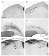
Retinal projections to the subcortical visual system in congenic albino and pigmented rats
- PMID: 16996223
- PMCID: PMC1876705
- DOI: 10.1016/j.neuroscience.2006.08.016
Retinal projections to the subcortical visual system in congenic albino and pigmented rats
Abstract
The primary visual pathway in albino mammals is characterized by an increased decussation of retinal ganglion cell axons at the optic chiasm and an enhanced contralateral projection to the dorsal lateral geniculate nucleus. In contrast to the primary visual pathway, little is known about the organization of retinal input to most nuclei of the subcortical visual system in albino mammals. The subcortical visual system is a large group of retinorecipient nuclei in the diencephalon and mesencephalon. These areas mediate a range of behaviors that include both circadian and acute responses to light. We used a congenic strain of albino and pigmented rats with a mutation at the c locus for albinism (Fischer 344-c/+; LaVail MM, Lawson NR (1986) Development of a congenic strain of pigmented and albino rats for light damage studies. Exp Eye Res 43:867-869) to quantitatively assess the effects of albinism on retinal projections to a number of subcortical visual nuclei including the ventral lateral hypothalamus (VLH), ventral lateral preoptic area (VLPO), olivary pretectal nucleus (OPN), posterior limitans (PLi), commissural pretectal area (CPA), intergeniculate leaflet (IGL), ventral lateral geniculate nucleus (vLGN) and superior colliculus (SC). Following eye injections of the neuroanatomical tracer cholera toxin-beta, the distribution of anterogradely transported label was measured. The retinal projection to the contralateral VLH, PLi, CPA and IGL was enhanced in albino rats. No significant differences were found between albino and pigmented rats in retinal input to the VLPO, OPN and vLGN. These findings raise the possibility that enhanced retinofugal projections to subcortical visual nuclei in albinos may underlie some light-mediated behaviors that differ between albino and pigmented mammals.
Figures


Similar articles
-
Retinal projections in the short-tailed fruit bat, Carollia perspicillata, as studied using the axonal transport of cholera toxin B subunit: Comparison with mouse.J Comp Neurol. 2015 Aug 15;523(12):1756-91. doi: 10.1002/cne.23723. Epub 2015 Jun 5. J Comp Neurol. 2015. PMID: 25503714
-
Interconnections among nuclei of the subcortical visual shell: the intergeniculate leaflet is a major constituent of the hamster subcortical visual system.J Comp Neurol. 1998 Jul 6;396(3):288-309. J Comp Neurol. 1998. PMID: 9624585
-
Retinal projections to the pretectum, accessory optic system and superior colliculus in pigmented and albino ferrets.Eur J Neurosci. 1993 May 1;5(5):486-500. doi: 10.1111/j.1460-9568.1993.tb00515.x. Eur J Neurosci. 1993. PMID: 8261124
-
Visual system of a naturally microphthalmic mammal: the blind mole rat, Spalax ehrenbergi.J Comp Neurol. 1993 Feb 15;328(3):313-50. doi: 10.1002/cne.903280302. J Comp Neurol. 1993. PMID: 8440785 Review.
-
The ventral lateral geniculate nucleus and the intergeniculate leaflet: interrelated structures in the visual and circadian systems.Neurosci Biobehav Rev. 1997 Sep;21(5):705-27. doi: 10.1016/s0149-7634(96)00019-x. Neurosci Biobehav Rev. 1997. PMID: 9353800 Review.
Cited by
-
Age-related changes in neurochemical components and retinal projections of rat intergeniculate leaflet.Age (Dordr). 2016 Feb;38(1):4. doi: 10.1007/s11357-015-9867-9. Epub 2015 Dec 30. Age (Dordr). 2016. PMID: 26718202 Free PMC article.
-
Longitudinal Assessments of Normal and Perilesional Tissues in Focal Brain Ischemia and Partial Optic Nerve Injury with Manganese-enhanced MRI.Sci Rep. 2017 Feb 23;7:43124. doi: 10.1038/srep43124. Sci Rep. 2017. PMID: 28230106 Free PMC article.
-
Imaging axonal transport in the rat visual pathway.Biomed Opt Express. 2013 Feb 1;4(2):364-86. doi: 10.1364/BOE.4.000364. Epub 2013 Jan 30. Biomed Opt Express. 2013. PMID: 23412846 Free PMC article.
-
Light-induced responses of slow oscillatory neurons of the rat olivary pretectal nucleus.PLoS One. 2012;7(3):e33083. doi: 10.1371/journal.pone.0033083. Epub 2012 Mar 12. PLoS One. 2012. PMID: 22427957 Free PMC article.
-
Retinal projections to the accessory optic system in pigmented and albino ferrets (Mustela putorius furo).Exp Brain Res. 2009 Dec;199(3-4):333-43. doi: 10.1007/s00221-008-1690-4. Exp Brain Res. 2009. PMID: 19139858
References
-
- Akerman CJ, Tolhurst DJ, Morgan JE, Baker GE, Thompson ID. Relay of visual information to the lateral geniculate nucleus and the visual cortex in albino ferrets. J Comp Neurol. 2003;461:217–235. - PubMed
-
- Albers FJ, Meek J, Nieuwenhuys R. Morphometric parameters of the superior colliculus of albino and pigmented rats. J Comp Neurol. 1988;274:357–370. - PubMed
-
- Balkema GW, Mangini NJ, Pinto LH, Vanable JW., Jr Visually evoked eye movements in mouse mutants and inbred strains. A screening report. Invest Ophthalmol Vis Sci. 1984;25:795–800. - PubMed
-
- Balkema GW. Elevated dark-adapted thresholds in albino rodents. Invest Ophthalmol Vis Sci. 1988;29:544–549. - PubMed
-
- Balkema GW, Drager UC. Impaired visual thresholds in hypopigmented animals. Vis Neurosci. 1991;6:577–585. - PubMed
Publication types
MeSH terms
Substances
Grants and funding
LinkOut - more resources
Full Text Sources
Research Materials
Miscellaneous

