

Pattern of hemodynamic impairment in multiple sclerosis: dynamic susceptibility contrast perfusion MR imaging at 3.0 T
- PMID: 16996280
- PMCID: PMC1752216
- DOI: 10.1016/j.neuroimage.2006.08.008
Pattern of hemodynamic impairment in multiple sclerosis: dynamic susceptibility contrast perfusion MR imaging at 3.0 T
Abstract
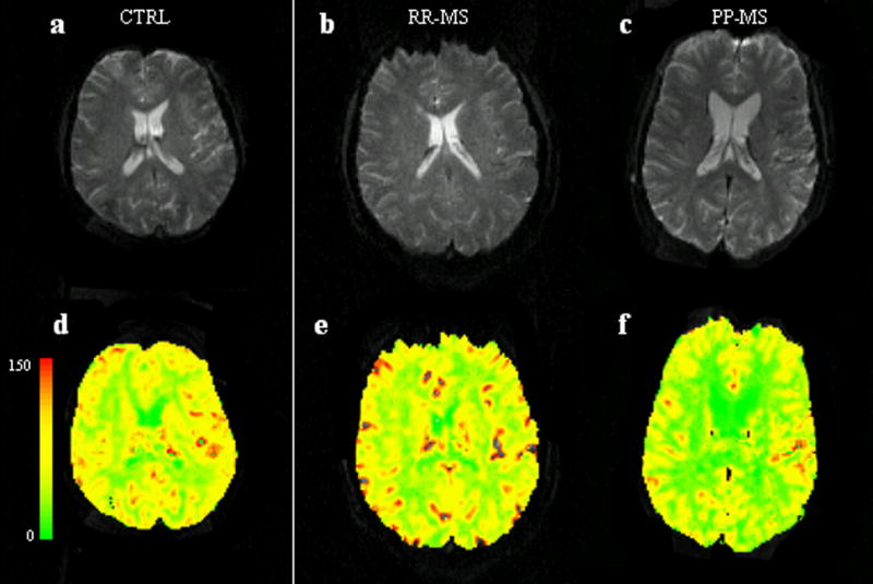
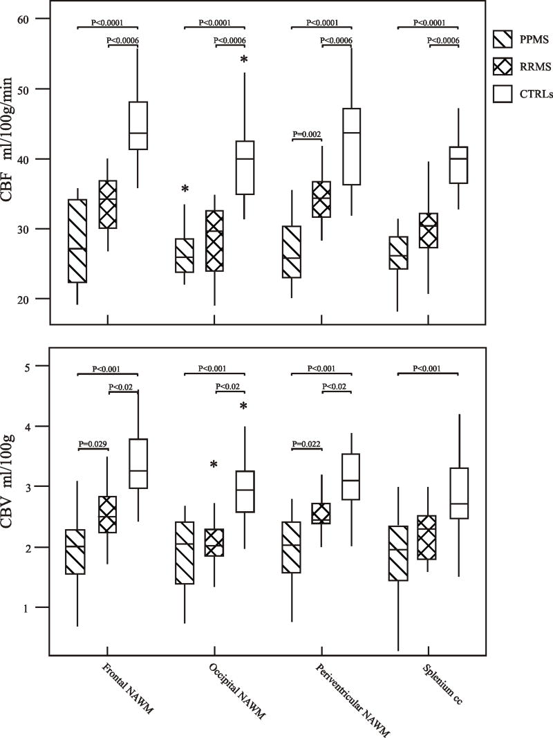
This study aimed to determine regional pattern of tissue perfusion in the normal-appearing white matter (NAWM) of patients with primary-progressive (PP), relapsing-remitting (RR) multiple sclerosis (MS) and healthy controls, and to investigate the association between perfusion abnormalities and clinical disability. Using dynamic susceptibility contrast (DSC) perfusion MRI at 3 T, we studied 22 patients with clinically definite MS, 11 with PP-MS and 11 with RR-MS and 11 age- and gender-matched healthy volunteers. The MRI protocol included axial dual-echo, dynamic susceptibility contrast enhanced (DSC) T2*-weighted and post-contrast T1-weighted images. Absolute cerebral blood flow (CBF), cerebral blood volume (CBV) and mean transit time (MTT) were measured in the periventricular, frontal, occipital NAWM and in the splenium of the corpus callosum. Compared to controls, CBF and CBV were significantly lower in all NAWM regions in both PP-MS patients (p values from <0.0001 to 0.001) and RR-MS (p values from <0.0001 to 0.020). Compared to RR-MS, PP-MS patients showed significantly lower CBF in the periventricular NAWM (p=0.002) and lower CBV in the periventricular and frontal NAWM (p values: 0.0029 and 0.022). EDSS was significantly correlated with the periventricular CBF (r=-0.48, p=0.0016) and with the periventricular and frontal CBV (r=-0.42, p=0.015; r=-0.35, p=0.038, respectively). This study suggests that the hemodynamic abnormalities of NAWM have clinical relevance in patients with MS. DSC perfusion MRI might provide a relevant objective measure of disease activity and treatment efficacy.
Figures
Similar articles
-
Microvascular abnormality in relapsing-remitting multiple sclerosis: perfusion MR imaging findings in normal-appearing white matter.Radiology. 2004 Jun;231(3):645-52. doi: 10.1148/radiol.2313030996. Radiology. 2004. PMID: 15163806
-
Dynamic Contrast-Enhanced Magnetic Resonance Imaging Suggests Normal Perfusion in Normal-Appearing White Matter in Multiple Sclerosis.Invest Radiol. 2017 Mar;52(3):135-141. doi: 10.1097/RLI.0000000000000320. Invest Radiol. 2017. PMID: 27548346
-
White matter hemodynamic abnormalities precede sub-cortical gray matter changes in multiple sclerosis.J Neurol Sci. 2009 Jul 15;282(1-2):28-33. doi: 10.1016/j.jns.2008.12.036. Epub 2009 Jan 31. J Neurol Sci. 2009. PMID: 19181347 Free PMC article.
-
Quantification of perfusion and permeability in multiple sclerosis: dynamic contrast-enhanced MRI in 3D at 3T.Invest Radiol. 2012 Apr;47(4):252-8. doi: 10.1097/RLI.0b013e31823bfc97. Invest Radiol. 2012. PMID: 22373532
-
Relationship between MRI perfusion and clinical severity in multiple sclerosis.Neural Regen Res. 2020 Apr;15(4):646-652. doi: 10.4103/1673-5374.266906. Neural Regen Res. 2020. PMID: 31638086 Free PMC article. Review.
Cited by
-
Microvascular changes in the macular and parafoveal areas of multiple sclerosis patients without optic neuritis.Sci Rep. 2022 Aug 3;12(1):13366. doi: 10.1038/s41598-022-17344-3. Sci Rep. 2022. PMID: 35922463 Free PMC article.
-
Hypoperfusion of brain parenchyma is associated with the severity of chronic cerebrospinal venous insufficiency in patients with multiple sclerosis: a cross-sectional preliminary report.BMC Med. 2011 Mar 7;9:22. doi: 10.1186/1741-7015-9-22. BMC Med. 2011. PMID: 21385345 Free PMC article.
-
A semi-automated measuring system of brain diffusion and perfusion magnetic resonance imaging abnormalities in patients with multiple sclerosis based on the integration of coregistration and tissue segmentation procedures.BMC Med Imaging. 2016 Jan 14;16:4. doi: 10.1186/s12880-016-0108-1. BMC Med Imaging. 2016. PMID: 26762399 Free PMC article.
-
Angiography with optical coherence tomography as a biomarker in multiple sclerosis.PLoS One. 2020 Dec 8;15(12):e0243236. doi: 10.1371/journal.pone.0243236. eCollection 2020. PLoS One. 2020. PMID: 33290417 Free PMC article.
-
Quantitative magnetic resonance imaging towards clinical application in multiple sclerosis.Brain. 2021 Jun 22;144(5):1296-1311. doi: 10.1093/brain/awab029. Brain. 2021. PMID: 33970206 Free PMC article. Review.
References
-
- Adams CW, Poston RN, Buk SJ, Sidhu YS, Vipond H. Inflammatory vasculitis in multiple sclerosis. J Neurol Sci. 1985;69:269–283. - PubMed
-
- Adams RA, Passino M, Sachs BD, Nuriel T, Akassoglou K. Fibrin mechanisms and functions in nervous system pathology. Mol Interv. 2004;4:163–176. - PubMed
-
- Axel L. Cerebral blood flow determination by rapid-sequence computed tomography: theoretical analysis. Radiology. 1980;137:679–686. - PubMed
-
- Bermel RA, Sharma J, Tjoa CW, Puli SR, Bakshi R. A semiautomated measure of whole-brain atrophy in multiple sclerosis. J Neurol Sci. 2003;208:57–65. - PubMed
Publication types
MeSH terms
Grants and funding
LinkOut - more resources
Full Text Sources
Other Literature Sources
Medical
Miscellaneous
