. 2007 Jan 15;301(2):417-31.
doi: 10.1016/j.ydbio.2006.08.047. Epub 2006 Aug 24.

Intermolecular interactions of homologs of germ plasm components in mammalian germ cells

Affiliations

Intermolecular interactions of homologs of germ plasm components in mammalian germ cells

Mark S Fox et al. Dev Biol. .

Abstract

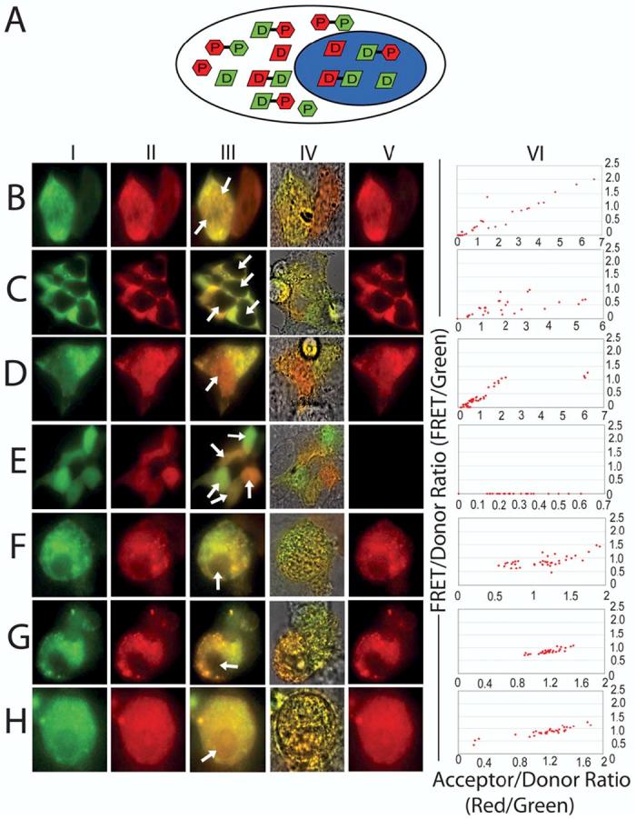
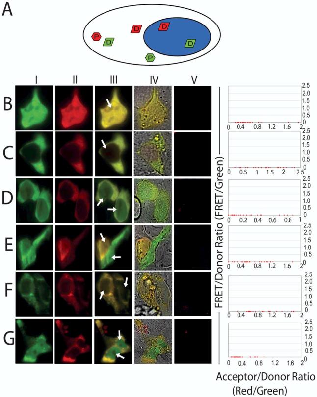
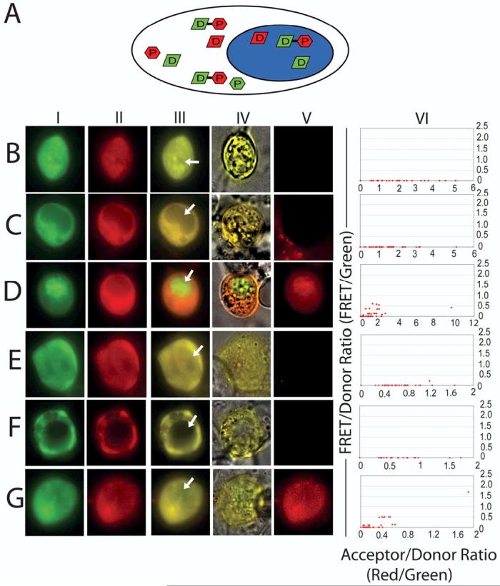
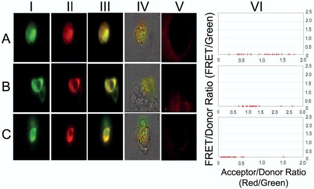
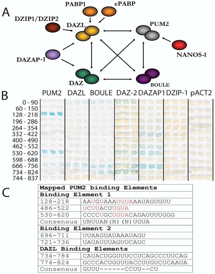
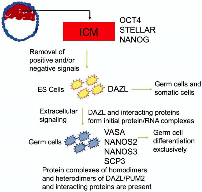
In some species such as flies, worms, frogs and fish, the key to forming and maintaining early germ cell populations is the assembly of germ plasm, microscopically distinct egg cytoplasm that is rich in RNAs, RNA-binding proteins and ribosomes. Cells which inherit germ plasm are destined for the germ cell lineage. In contrast, in mammals, germ cells are formed and maintained later in development as a result of inductive signaling from one embryonic cell type to another. Research advances, using complementary approaches, including identification of key signaling factors that act during the initial stages of germ cell development, differentiation of germ cells in vitro from mouse and human embryonic stem cells and the demonstration that homologs of germ plasm components are conserved in mammals, have shed light on key elements in the early development of mammalian germ cells. Here, we use FRET (Fluorescence Resonance Energy Transfer) to demonstrate that living mammalian germ cells possess specific RNA/protein complexes that contain germ plasm homologs, beginning in the earliest stages of development examined. Moreover, we demonstrate that, although both human and mouse germ cells and embryonic stem cells express the same proteins, germ cell-specific protein/protein interactions distinguish germ cells from precursor embryonic stem cells in vitro; interactions also determine sub-cellular localization of complex components. Finally, we suggest that assembly of similar protein complexes may be central to differentiation of diverse cell lineages and provide useful diagnostic tools for isolation of specific cell types from the assorted types differentiated from embryonic stem cells.


Figures

Fig 1
Fig 1
Schematic of methods used to analyze protein complexes in germ cells. As shown, mouse testis extract was prepared in the presence of either RNAsin or RNase A in both physiological and high salt conditions (500 mM NaCl). To examine whether DAZL and PUM2 participate in the formation of one or several complexes in vivo, cytoplasmic extracts were then subjected to size exclusion chromatography and components of the eluants were analyzed by Western blotting. The relative contributions of protein-RNA versus protein-protein interactions can be approximated via comparison of protein complex profiles from different treatments.
Fig. 2
Fig. 2
Protein complexes revealed by size exclusion chromatography. (A) Cytoplasmic lysate from mouse testis treated with RNAsin (I) or RNAse A (II) in physiological salt concentrations (150mM NaCl) was fractionated by size exclusion chromatography; absorbance readings at 280nm for each 0.5 ml-fraction were plotted. (B) The presence of DAZL, PUM2, NANOS1, VASA, BOULE, DAZAP1, and Ribosomal P proteins was visualized by Western blotting of proteins in extracts treated with either RNAsin (I) or RNase A (II) in physiological salt concentrations (150mM NaCl). The position of molecular-weight makers separated in parallel on the same column is indicated below the Western blot. (C) Cytoplasmic lysate from mouse testis treated with RNAsin (I) or RNAse A (II) was fractionated by size exclusion chromatography in high salt concentrations (500mM NaCl); absorbance readings at 280nm for each fraction were plotted. (D) The presence of DAZL, PUM2, NANOS1, VASA, BOULE, DAZAP1, and Ribosomal P proteins was visualized by Western blotting of proteins in extracts treated with either RNAsin (I) or RNase A (II) in high salt concentrations (500mM NaCl). The position of molecular-weight markers separated in parallel on the same column is indicated below the Western blot. Molecular weight markers were thyroglobulin (670 KD), gamma-globulin (158 KD), ovalbumin (44 KD), and Vitamin B12 (1.3 KD).
Fig. 3
Fig. 3
DAZL and PUM2 proteins in living postnatal mouse germ cells. (A) Reference diagram of a postnatal germ cell with the nucleus colored in blue. Cellular distribution of DAZL and PUM2 subunits are depicted by the letters D and P respectively; D-D and P-P correspond to homodimers while D-P is used to depict DAZL/PUM2 complexes. (B) eGFP-DAZL interaction with RFP-DAZL in spermatogonial stem cells. (C) eGFP-PUM2 interaction with RFP-PUM2 in spermatogonial stem cells. (D) eGFP-DAZL interaction with RFP-PUM2 in spermatogonial stem cells. (E) Negative control eGFP interaction with RFP in spermatogonial stem cells. (F) eGFP-DAZL interaction with RFP-DAZL in primary germ cells from day 3 mice. (G) eGFP-PUM2 interaction with RFP-PUM2 in primary germ cells from day 3 mice. (H) eGFP-DAZL interaction with RFP-PUM2 in primary germ cells from day 3 mice. Panels I - VI in each row as follows: (I) A reference image showing expression of eGFP fusion constructs throughout the stem cell. (II) A second digital image obtained from the same plane showing expression of RFP fusion construct. (III) Colocalization of panels I and II depicted in yellow. (IV) Phase contrast overlay of Panels I and II. (V) FRET detected by stimulating the cells with blue light and using the acceptor filter for the RFP protein. (VI) FRET/donor ratios as graphed as a function of acceptor/donor ratios.
Fig. 4
Fig. 4
DAZL and PUM2 proteins in living embryonic stem cells. (A) Reference diagram of a embryonic stem cell with the nucleus colored in blue. Cellular distribution of DAZL and PUM2 subunits are depicted by the letters D and P respectively. (B) eGFP-DAZL interaction with RFP-DAZL in mouse embryonic stem cells. (C) eGFP-PUM2 interaction with RFP-PUM2 in mouse embryonic stem cells. (D) eGFP-DAZL interaction with RFP-PUM2 in mouse embryonic stem cells. (E) eGFP-DAZL interaction with RFP-DAZL in human embryonic stem cells. (F) eGFP-PUM2 interaction with RFP-PUM2 in human embryonic stem cells. (G) eGFP-DAZL interaction with RFP-PUM2 in human embryonic stem cells. Panels I - VI in each row as follows: (I) A reference image showing expression of eGFP fusion constructs throughout the stem cell. (II) A second digital image obtained from the same plane showing expression of RFP fusion construct. (III) Colocalization of panels I and II depicted in yellow. (IV) Phase contrast overlay of Panels I and II. (V) FRET detected by stimulating the cells with blue light and using the acceptor filter for the RFP protein. (VI) FRET/donor ratios as graphed as a function of acceptor/donor ratios.
Fig. 5
Fig. 5
DAZL and PUM2 proteins in living mouse embryonic germ cells from day 8.5 and 12.5 embryos. (A) Reference diagram of a postnatal germ cell with the nucleus colored in blue. Cellular distribution of DAZL and PUM2 subunits are depicted by the letters D and P respectively while D-P is used to depict DAZL/PUM2 complexes. (BA) eGFP-DAZL interaction with RFP-DAZL in embryonic germ cells from day 8.5. (C) eGFP-PUM2 interaction with RFP-PUM2 in embryonic germ cells from day 8.5. (D) eGFP-DAZL interaction with RFP-PUM2 in embryonic germ cells from day 8.5. (E) eGFP-DAZL interaction with RFP-DAZL in embryonic germ cells from day 12.5. (F) eGFP-PUM2 interaction with RFP-PUM2 in embryonic germ cells from day 12.5. (G) eGFP-DAZL interaction with RFP-PUM2 in embryonic germ cells from day 12.5. Panels I - VI in each row as follows: (I) A reference image showing expression of eGFP fusion constructs throughout the stem cell. (II) A second digital image obtained from the same plane showing expression of RFP fusion construct. (III) Colocalization of panels I and II depicted in yellow. (IV) Phase contrast overlay of Panels I and II. (V) FRET detected by stimulating the cells with blue light and using the acceptor filter for the RFP protein. (VI) FRET/donor ratios as graphed as a function of acceptor/donor ratios. (B) eGFP-DAZL interaction with RFP-DAZL in embryonic germ cells from day 8.5. (C) eGFP-PUM2 interaction with RFP-PUM2 in embryonic germ cells from day 8.5. (D) eGFP-DAZL interaction with RFP-PUM2 in embryonic germ cells from day 8.5. (E) eGFP-DAZL interaction with RFP-DAZL in embryonic germ cells from day 12.5. (F) eGFP-PUM2 interaction with RFP-PUM2 in embryonic germ cells from day 12.5. (G) eGFP-DAZL interaction with RFP-PUM2 in embryonic germ cells from day 12.5. Panels I - VI in each row as follows: (I) A reference image showing expression of eGFP fusion constructs throughout the stem cell. (II) A second digital image obtained from the same plane showing expression of RFP fusion construct. (III) Colocalization of panels I and II depicted in yellow with the location of the nucleus indicated by an arrow. (IV) Phase contrast overlay of Panels I and II. (V) FRET detected by stimulating the cells with blue light and using the acceptor filter for the RFP protein. (VI) FRET/donor ratios as graphed as a function of acceptor/donor ratios.
Fig. 6
Fig. 6
DAZL and PUM2 proteins in living mouse Leydig cells. (A) eGFP-DAZL interaction with RFP-DAZL. (B) eGFP-PUM2 interaction with RFP-PUM2. (C) eGFP-DAZL interaction with RFP-PUM2. Panels I - VI in each row as follows: (I) A reference image showing expression of eGFP fusion constructs throughout the stem cell. (II) A second digital image obtained from the same plane showing expression of RFP fusion construct. (III) Colocalization of panels I and II depicted in yellow. (IV) Phase contrast overlay of Panels I and II. (V) FRET detected by stimulating the cells with blue light and using the acceptor filter for the RFP protein. (VI) FRET/donor ratios as graphed as a function of acceptor/donor ratios.
Fig. 7
Fig. 7
Multiple interacting proteins may bind the same RNA. (A) Diagram illustrating known protein-protein interactions with DAZL and PUM2. In addition to interacting with each other, DAZL and PUM2 can interact with several other RNA-binding proteins that also, in turn can interact with each other. (B) Binding of proteins to the 3’UTR of an mRNA transcript with DAZL and PUM2 consensus binding sequences. Fragments are numbered from 0 to 837, corresponding to the stop codon and polyadenylated tail of the SDAD1 transcript, respectively. Blue color indicates binding of the RNA sequence by the indicated proteins in the yeast three-hybrid assay. Binding was detected for fragments 0-90, 128-218, 332-422, 462-552, 530-620, 666-756 and 734-837 by several proteins, but not DZIP1. (C) Specificity of PUM2 and DAZL binding to the 3’ UTR of SDAD1. Using yeast three hybrid and electrophoretic mobility shift (gel shift) assays, PUM2 was shown to recognize five regions within the 3’UTR of the transcript SDAD1that were divided into two separate binding elements. The nucleotides required for binding for PUM2 binding element 1 are highlighted in red and a consensus binding element was also defined. Similarly, two regions were mapped for DAZL and a consensus sequence determined by alignment with all four regions bound by DAZL is shown below (Fox et al., 2005).
Fig. 8
Fig. 8
Model of germ cell differentiation based on these studies and others. See text for further explanation.

Similar articles

Cited by

References

    1. Abeyta M, Clark AT, Rodriguez R, Bodnar M, Reijo Pera RA, Firpo M. Unique gene expression signatures of independently-derived human embryonic stem cell lines. Hum Mol Genet. 2004;13:601–608. - PubMed
    1. Asaoka-Taguchi M, Yamada M, Nakamura A, Hanyu K, Kobayashi S. Maternal Pumilio acts together with Nanos in germline development in Drosophila embryos. Nature Cell Biol. 1999;1:431–7. - PubMed
    1. Buehr M, Smith A. Genesis of embryonic stem cells. Philos Trans R Soc Lond B Biol Sci. 2003;358:1397–1402. - PMC - PubMed
    1. Campbell R, Tour O, Palmer A, Steinbach P, Baird G, Zacharias D, Tsien R. A monomeric red fluorescent protein. Proc Natl Acad Sci. 2002;99:7877–7882. - PMC - PubMed
    1. Castrillon DH, Quade BJ, Wang TY, Quigley C, Crum CP. The human VASA gene is specifically expressed in the germ cell lineage. Proc Natl Acad Sci. 2000;97:9585–9590. - PMC - PubMed

Publication types

Substances

LinkOut - more resources