

Dyskinesis in Chagasic myocardium: centerline analysis of wall motion using cardiac-gated magnetic resonance images of mice
- PMID: 16997075
- PMCID: PMC2654323
- DOI: 10.1016/j.mri.2006.04.001
Dyskinesis in Chagasic myocardium: centerline analysis of wall motion using cardiac-gated magnetic resonance images of mice
Abstract
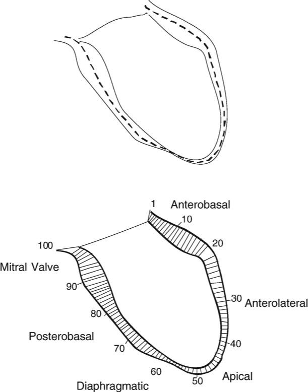
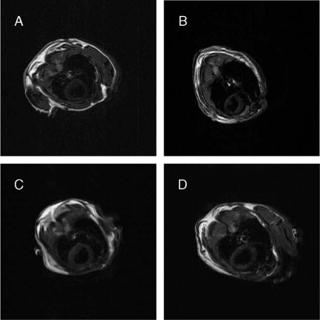
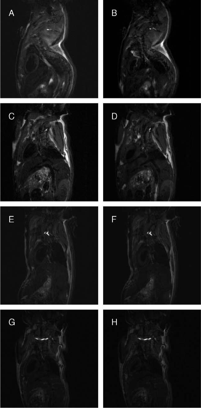
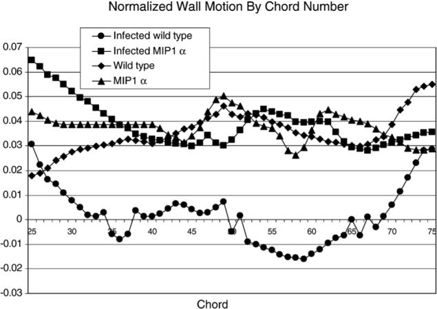
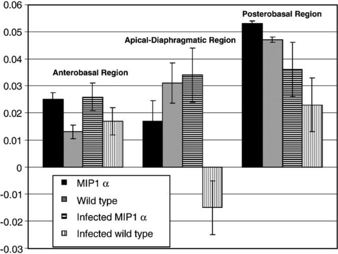
We report on the use of centerline analysis of cardiac-gated magnetic resonance images to measure wall motion abnormalities in mice infected with Trypanosoma cruzi. To our knowledge, this is the first report of segmental wall motion abnormalities in an animal model of Chagas' disease. Chagas' disease patients with severe cardiac involvement exhibit mild hypokinesis in an extensive region of the left ventricle and dyskinesis in the apical region. We observed dyskinetic segments in a similar region of the hearts of infected wild-type mice. Dyskinesis was not observed in infected mice lacking macrophage inflammatory protein-1alpha, a chemokine that may play an important role in the cardiac remodeling that is normally observed in mouse models of Chagas' disease and in human patients. This study aimed to demonstrate the utility of cardiac-gated magnetic resonance imaging and centerline analysis as a straightforward method for monitoring regional left ventricular wall motion in transgenic and/or diseased mice.
Figures
Similar articles
-
Improved Biomarker and Imaging Analysis for Characterizing Progressive Cardiac Fibrosis in a Mouse Model of Chronic Chagasic Cardiomyopathy.J Am Heart Assoc. 2019 Nov 19;8(22):e013365. doi: 10.1161/JAHA.119.013365. Epub 2019 Nov 13. J Am Heart Assoc. 2019. PMID: 31718442 Free PMC article.
-
Histopathological Correlates of Global and Segmental Left Ventricular Systolic Dysfunction in Experimental Chronic Chagas Cardiomyopathy.J Am Heart Assoc. 2016 Jan 21;5(1):e002786. doi: 10.1161/JAHA.115.002786. J Am Heart Assoc. 2016. PMID: 26796255 Free PMC article.
-
Myocardial tissue characterization in Chagas' heart disease by cardiovascular magnetic resonance.J Cardiovasc Magn Reson. 2015 Nov 18;17:97. doi: 10.1186/s12968-015-0200-7. J Cardiovasc Magn Reson. 2015. PMID: 26581396 Free PMC article.
-
Magnetic resonance imaging in experimental Chagas disease: a brief review of the utility of the method for monitoring right ventricular chamber dilatation.Parasitol Res. 2005 Sep;97(2):87-90. doi: 10.1007/s00436-005-1409-4. Epub 2005 Jun 29. Parasitol Res. 2005. PMID: 15986245 Review.
-
[Chagasic myocardiopathy: historical perspective].Medicina (B Aires). 1999;59 Suppl 2:25-40. Medicina (B Aires). 1999. PMID: 10668240 Review. Spanish.
Cited by
-
Chagas heart disease: report on recent developments.Cardiol Rev. 2012 Mar-Apr;20(2):53-65. doi: 10.1097/CRD.0b013e31823efde2. Cardiol Rev. 2012. PMID: 22293860 Free PMC article. Review.
-
NADPH phagocyte oxidase knockout mice control Trypanosoma cruzi proliferation, but develop circulatory collapse and succumb to infection.PLoS Negl Trop Dis. 2012;6(2):e1492. doi: 10.1371/journal.pntd.0001492. Epub 2012 Feb 14. PLoS Negl Trop Dis. 2012. PMID: 22348160 Free PMC article.
-
Advances in imaging of animal models of Chagas disease.Adv Parasitol. 2011;75:193-208. doi: 10.1016/B978-0-12-385863-4.00009-5. Adv Parasitol. 2011. PMID: 21820557 Free PMC article.
-
Pathogenesis of Chagas disease: time to move on.Front Biosci (Elite Ed). 2012 Jan 1;4(5):1743-58. doi: 10.2741/495. Front Biosci (Elite Ed). 2012. PMID: 22201990 Free PMC article.
-
A robust computational framework for estimating 3D Bi-Atrial chamber wall thickness.Comput Biol Med. 2019 Nov;114:103444. doi: 10.1016/j.compbiomed.2019.103444. Epub 2019 Sep 12. Comput Biol Med. 2019. PMID: 31542646 Free PMC article.
References
-
- Henson RE, Song SK, Pastorek JS, Ackerman JJH, Lorenz CH. Left ventricular torsion is equal in mice and humans. Am J Physiol. 1999;278:H1117–23. - PubMed
-
- Vignon P, Weinert L, Mor-Avi V, Spencer KT, Bednarz J, Lang RM. Quantitative assessment of regional right ventricular function with color kinesis. Am J Respir Crit Care Med. 1999;159:1949–59. - PubMed
-
- Koch R, Lang RM, Garcia MJ, Weinert L, Bednarz J, Korcarz C, et al. Objective evaluation of regional left ventricular wall motion during dobutamine stress echocardiographic studies using segmental analysis of color kinesis images. J Am Coll Cardiol. 1999;34:409–19. - PubMed
-
- Scherrer-Crosbie M, Steudel W, Hunziker PR, Liel-Cohen N, Ullrich WM, Zapol WM, et al. Three-dimensional echocardiographic assessment of left ventricular wall motion abnormalities in mouse myocardial infarction. J Am Soc Echocardiogr. 1999;12:834–40. - PubMed
-
- Hubka M, Lipiecki J, Bolson EL, Martin RW, Munt B, Maza SR, et al. Three-dimensional echocardiographic measurement of left ventricular wall thickness: in vitro and in vivo validation. J Am Soc Echocardiogr. 2002;15:129–35. - PubMed
Publication types
MeSH terms
Grants and funding
LinkOut - more resources
Full Text Sources
