. 2006 Dec;188(23):8252-8.
doi: 10.1128/JB.01213-06. Epub 2006 Sep 22.

Multiple GTPases participate in the assembly of the large ribosomal subunit in Bacillus subtilis

Affiliations

Multiple GTPases participate in the assembly of the large ribosomal subunit in Bacillus subtilis

Laura Schaefer et al. J Bacteriol. 2006 Dec.

Abstract

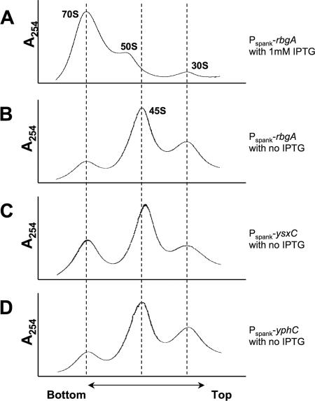
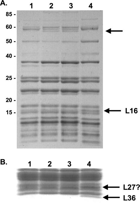
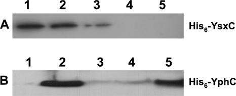
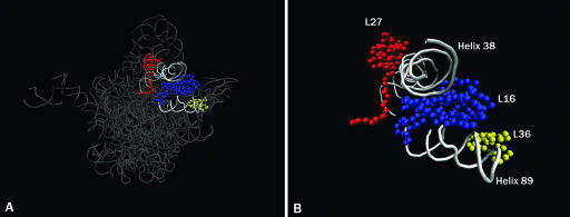
GTPases have been demonstrated to be necessary for the proper assembly of the ribosome in bacteria and eukaryotes. Here, we show that the essential GTPases YphC and YsxC are required for large ribosomal subunit biogenesis in Bacillus subtilis. Sucrose density gradient centrifugation of large ribosomal subunits isolated from YphC-depleted cells and YsxC-depleted cells indicates that they are similar to the 45S intermediate previously identified in RbgA-depleted cells. The sedimentation of the large-subunit intermediate isolated from YphC-depleted cells was identical to the intermediate found in RbgA-depleted cells, while the intermediate isolated from YsxC-depleted cells sedimented slightly slower than 45S, suggesting that it is a novel intermediate. Analysis of the protein composition of the large-subunit intermediates isolated from either YphC-depleted cells or YsxC-depleted cells indicated that L16 and L36 are missing. Purified YphC and YsxC are able to interact with the ribosome in vitro, supporting a direct role for these two proteins in the assembly of the 50S subunit. Our results indicate that, as has been demonstrated for Saccharomyces cerevisiae ribosome biogenesis, bacterial 50S ribosome assembly requires the function of multiple essential GTPases.


Figures

FIG. 1.
FIG. 1.
Growth curves of strains depleted of YsxC or YphC. Strains RB260 (Pspank-yxsC) and RB290 (Pspank-yphC) were grown for several generations in the presence of 1 mM IPTG or without an inducer. When the cultures reached on OD600 of 1.0, they were diluted back into prewarmed LB medium. Circles, RB260 grown in the presence of 1 mM IPTG (YsxC+); diamonds, RB290 grown in the presence of 1 mM IPTG (YphC+); squares, RB260 grown in the absence of IPTG (YsxC); ×, RB290 grown in the absence of IPTG (YphC).
FIG. 2.
FIG. 2.
Ribosome biogenesis defects in YsxC- and YphC-depleted cells. Ribosome profiles were generated by centrifugation of lysates through a 10 to 25% sucrose gradient. (A) Wild-type ribosome profile from the RB301 strains that is fully induced for RbgA expression in the presence of 1 mM IPTG. Ribosome profiles of cells depleted of (B) RbgA, (C) YsxC, and (D) YphC are also shown. Dashed lines indicate the migration of 70S subunits, the 45S intermediate, and mature 30S subunits.
FIG. 3.
FIG. 3.
Analysis of the protein content of the ribosomal intermediates isolated from YsxC- and YphC-depleted cells. (A) A 12% SDS-PAGE gel containing purified large-subunit intermediates. Molecular masses (kDa) are indicated on the left side of the gel. Lane 1, 44.5S intermediate isolated from YsxC-depleted cells; lane 2, 45S intermediate isolated from YphC-depleted cells; lane 3, 45S intermediate isolated from RbgA-depleted cells; lane 4, mature 50S subunits isolated from wild-type cells. The bottom arrow indicates where L16 is present in the 50S subunit but absent from the intermediates. The top arrow indicates a region of the gel where the 44.5S complex differs from the 45S intermediates. The identities of the proteins are not yet known, although their sizes indicate that they cannot be ribosomal proteins. (B) A 16% SDS-PAGE gel containing purified large-subunit intermediates. Only the region of the gel at 12 kDa and smaller is shown. Lane 1, 44.5S complex from YsxC-depleted cells; lane 2, 45S complex from YphC-depleted cells; lane 3, 45S complex from RbgA-depleted cells; lane 4, mature 50S subunits from wild-type cells. Arrows indicate the positions of L36 and possibly L27, found only in the mature 50S subunit.
FIG. 4.
FIG. 4.
YsxC and YphC directly interact with the ribosome. (A) Analysis of His6-YsxC by Western blotting of the pellets obtained from centrifugation on a 10% sucrose cushion of the mixtures of His6-YsxC and ribosome incubated in the presence of 1 mM GDP (lane 1), His6-YsxC and ribosome incubated in the presence of 1 mM GTP (lane 2), His6-YsxC and ribosome incubated without any added nucleotide (lane 3), ribosome alone (lane 4), and His6-YsxC alone (lane 5). His6-YsxC incubated with 1 mM GTP or 1 mM GDP gave a signal that was similar to that of His6-YsxC alone (lane 1), indicating that His6-YsxC does not significantly precipitate or aggregate in the presence of nucleotides (data not shown). (B) Analysis of His6-YphC by Western blotting of the pellets obtained from centrifugation on a 10% sucrose cushion of the mixtures of His6-YphC alone (lane 1), His6-YphC and ribosome (lane 2), His6-YphC and ribosome in presence of 2 mM GTP (lane 3), His6-YphC and ribosome in presence of 2 mM GDP (lane 4), and His6-YphC and ribosome in presence of 2 mM GMPPNP (lane 5). His6-YphC incubated with 2 mM GTP gave a signal that was similar to that of His6-YphC alone (lane 1), indicating that His6-YphC does not significantly precipitate or aggregate in the presence of nucleotides (data not shown).
FIG. 5.
FIG. 5.
Location of L16, L27, and L36 in the 50S subunit. (A) Crown view representation of the 50S subunit from Deinococcus radiodurans (Protein Data Bank accession number 1NKW). The locations of L16 (blue), L27 (red), and L36 (yellow) in the 50S subunit are shown. The rRNA is dark gray; helix 89 and helix 38 are shown in light gray. (B) Enlarged view of L16, L27, and L36 in the ribosome. Colors are the same as in A. Figures were generated using VMD software.

Similar articles

Cited by

References

    1. Anand, B., S. K. Verma, and B. Prakash. 2006. Structural stabilization of GTP-binding domains in circularly permuted GTPases: implications for RNA binding. Nucleic Acids Res. 34:2196-2205. - PMC - PubMed
    1. Arigoni, F., F. Talabot, M. Peitsch, M. D. Edgerton, E. Meldrum, E. Allet, R. Fish, T. Jamotte, M. L. Curchod, and H. Loferer. 1998. A genome-based approach for the identification of essential bacterial genes. Nat. Biotechnol. 16:851-856. - PubMed
    1. Ban, N., P. Nissen, J. Hansen, P. B. Moore, and T. A. Steitz. 2000. The complete atomic structure of the large ribosomal subunit at 2.4 A resolution. Science 289:905-920. - PubMed
    1. Bashan, A., I. Agmon, R. Zarivach, F. Schluenzen, J. Harms, R. Berisio, H. Bartels, F. Franceschi, T. Auerbach, H. A. Hansen, E. Kossoy, M. Kessler, and A. Yonath. 2003. Structural basis of the ribosomal machinery for peptide bond formation, translocation, and nascent chain progression. Mol. Cell 11:91-102. - PubMed
    1. Britton, R. A., B. S. Powell, S. Dasgupta, Q. Sun, W. Margolin, J. R. Lupski, and D. L. Court. 1998. Cell cycle arrest in Era GTPase mutants: a potential growth rate-regulated checkpoint in Escherichia coli. Mol. Microbiol. 27:739-750. - PubMed

Publication types

MeSH terms

LinkOut - more resources