. 2006 Oct;18(10):2517-30.
doi: 10.1105/tpc.106.045484. Epub 2006 Sep 22.

PDV1 and PDV2 mediate recruitment of the dynamin-related protein ARC5 to the plastid division site

Affiliations

PDV1 and PDV2 mediate recruitment of the dynamin-related protein ARC5 to the plastid division site

Shin-ya Miyagishima et al. Plant Cell. 2006 Oct.

Abstract

During plastid division, the dynamin-related protein ACCUMULATION AND REPLICATION OF CHLOROPLASTS5 (ARC5) is recruited from the cytosol to the surface of the outer chloroplast envelope membrane. In Arabidopsis thaliana arc5 mutants, chloroplasts arrest during division site constriction. Analysis of mutants similar to arc5 along with map-based cloning identified PLASTID DIVISION1 (PDV1), an integral outer envelope membrane protein, and its homolog PDV2 as components of the plastid division machinery. Similar to ARC5, PDV1 localized to a discontinuous ring at the division site in wild-type plants. The midplastid PDV1 ring formed in arc5 mutants and the ARC5 ring formed in pdv1 and pdv2 mutants, but not in pdv1 pdv2. Stromal FtsZ ring assembly occurred in pdv1, pdv2, and pdv1 pdv2, as it does in arc5. Topological analysis showed that the large N-terminal region of PDV1 upstream of the transmembrane helix bearing a putative coiled-coil domain is exposed to the cytosol. Mutation of the conserved PDV1 C-terminal Gly residue did not block PDV1 insertion into the outer envelope membrane but did abolish its localization to the division site. Our results indicate that plastid division involves the stepwise localization of FtsZ, PDV1, and ARC5 at the division site and that PDV1 and PDV2 together mediate the recruitment of ARC5 to the midplastid constriction at a late stage of division.


Figures

Figure 1.
Figure 1.
Phenotypes of arc5, pdv1, pdv2, and pdv1 pdv2 Mutants, and Complementation of the Phenotypes Conferred by pdv1. Chloroplasts in leaf mesophyll cells were observed by Nomarski optics, and a single cell is shown for each line. All mutants are Col-0 ecotype. Bar in (J) = 10 μm. (A) and (B) Wild-type Col-0 (A) and arc5-2 (SAIL_71D11) (B) for comparison. (C) to (F) Mutant phenotypes of pdv1-1 (C) and pdv1-2 (D) were complemented by a wild-type PDV1 transgene (pdv1-1 [E] and pdv1-2 [F]). (G) to (I) The wild type (G) and heterozygous (H) and homozygous (I) segregants of pdv2-1. (J) pdv1-1 pdv2-1. (K) Mutation sites of pdv1-1 and pdv1-2, and positions of T-DNA insertion of pdv2-1 and pdv2-2. Exons are depicted as rectangles, and nucleotide numbers from the annotated start codon are shown above the genes. nt, nucleotides. (L) Statistical comparison of the number of chloroplasts per mesophyll cell. Gray bars show mean numbers of chloroplasts, and error bars represent sd. n = 40 for the wild type and pdv2-1 heterozygous segregants; n = 80 for all others.
Figure 2.
Figure 2.
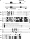
Predicted Structures of PDV1 and PDV2 Proteins, and Sequence Alignment of PDV1, PDV2, and Homologous Proteins. (A) Predicted structures of PDV1 and PDV2 proteins. Putative coiled-coil domains and transmembrane domains are depicted as closed rectangles, and their positions within the amino acid sequences are indicated. Black bars below the diagrams delineate the regions that are relatively well conserved among PDV1, PDV2, and related proteins based on the sequence alignment. aa, amino acids. (B) Sequence alignment of PDV1, PDV2, and related proteins. Black shading indicates residues identical in five or more sequences. The numbers of amino acids from the N terminus of Arabidopsis PDV1 are indicated above the alignment. A. th., Arabidopsis thaliana; rice, Oryza sativa; moss, Physcomitrella patens. Accession numbers for each sequence are listed in Methods. The sequences were aligned with ClustalW (Thompson et al., 1994).
Figure 3.
Figure 3.
Expression and Rough Fractionation of GFP-PDV1. Immunoblot analyses using anti-GFP antibody. Homogenates of flower buds from pdv1-1 plants expressing a PDV1 transgene (lane 1) and a GFP-PDV1 transgene (lane 2) were blotted. The homogenate containing GFP-PDV1 (Total; lane 3) was centrifuged at 20,000g to sediment the low-speed pellet fraction (LSP; lane 4). The supernatant fraction was fractionated further into the high-speed pellet (HSP; lane 5) and supernatant (S; lane 6) fractions at 100,000g. The low-speed pellet fraction was treated with 0.1 M sodium carbonate (lanes 7 and 8) or 1% Nonidet P-40 (NP-40; lanes 9 and 10), and then soluble (S; lanes 8 and 10) and insoluble (P; lanes 7 and 9) fractions were separated at 100,000g.
Figure 4.
Figure 4.
Localization of GFP-PDV1 and GFP-ARC5. A GFP-PDV1 transgene was expressed in pdv1-1 plants, and GFP fluorescence was observed by fluorescence microscopy. The fluorescence signal of GFP is green, and the autofluorescence of chlorophyll is red. The dim green fluorescence in (B) is background, because similar fluorescence was observed without the GFP-PDV1 transgene. Expression of the GFP-PDV1 transgene complemented the mutant phenotype of pdv1-1. Bars in (A), (B), and (J) = 5 μm. (C) to (J) are shown at the same magnification. (A) Chloroplasts in mesophyll cells of a young leaf expressing GFP-PDV1. (B) Plastids in fringes of petals from flower buds expressing GFP-PDV1. (C) to (F) Comparison of GFP-PDV1 localization among chloroplasts at various stages of division. (G) to (J) Comparison of GFP-ARC5 localization among chloroplasts at various stages of division.
Figure 5.
Figure 5.
Localization of FtsZ in arc5, pdv1, pdv2, and pdv1 pdv2 Mutants. Localization of FtsZ in mesophyll cells of young leaves was observed by immunofluorescence microscopy using anti-At FtsZ2-1 antibodies. The strong fluorescence is the GFP signal, and the dim fluorescence is the red autofluorescence of chlorophyll. (A) Wild type. (B) to (E) arc5-2 (B), pdv1-1 (C), pdv2-1 (D), and pdv1 pdv2 (E) mutants.
Figure 6.
Figure 6.
Localization of GFP-PDV1 in the arc5 Mutant, and Localization of GFP-ARC5 in pdv1, pdv2, and pdv1 pdv2 Mutants. (A) to (G) The strong fluorescence is the GFP signal, and the dim fluorescence is the red autofluorescence of chlorophyll. Mesophyll cells of young leaves were observed by fluorescence microscopy. Localization of GFP-PDV1 expressed in pdv1-1 (A) and arc5-1 (B) plants. Localization of GFP-ARC5 expressed in wild-type (C), pdv1-1 (D), pdv1-2 (E), pdv2-1 (F), and pdv1 pdv2 (G) plants. Bar in (G) = 5 μm. (H) Immunoblotting of extracts from young leaves showing the expression of GFP-ARC5 in pdv1 pdv2 mutant. Blots containing extracts from wild-type (lane 1) and pdv1 pdv2 (lane 2) plants expressing GFP-ARC5 were probed with anti-GFP antibody.
Figure 7.
Figure 7.
In Vitro Chloroplast Import, Protease Protection, and Fractionation Assay of PDV1. 35S-labeled PDV1, the integral outer envelope protein OEP14, the precursor of truncated inner envelope protein Tic110-110N (110-110N), and the precursor of the stroma-localized small subunit of ribulose-1,5-bis-phosphate carboxylase/oxygenase (SS) were synthesized in vitro. In vitro–translated proteins were incubated with isolated pea chloroplasts in the presence of 4.0 mM Mg-ATP for 30 min at room temperature. Chloroplasts were recovered by sedimentation through 40% (v/v) Percoll. p and m indicate precursor and mature proteins for 110-110N and SS, respectively. (A) The recovered intact chloroplasts were incubated without (−; lanes 2 and 3) or with (+; lanes 4 and 5) thermolysin for 30 min at 4°C. Intact chloroplasts were again recovered by centrifugation through 40% (v/v) Percoll and fractionated into total membrane (P; lanes 2 and 4) and soluble (S; lanes 3 and 5) fractions. TP represents 10% of translated product added to a single import assay (lane 1). (B) The recovered intact chloroplasts were lysed and fractionated into total membrane (P) and soluble (S) fractions (lanes 7 and 8). A portion of the membranes recovered in lane 7 for each import reaction was subsequently extracted with 0.1 M sodium carbonate, pH 11.5 (lanes 9 and 10), or with 1% Nonidet P-40 (lanes 11 and 12) for 1 h on ice. After the treatments, membranes (P) and extractable soluble proteins (S) were recovered and analyzed by SDS-PAGE and fluorography. TP represents 10% of translated product added to a single import assay (lane 6).
Figure 8.
Figure 8.
Effect of the C-Terminal Mutation of PDV1 (pdv1-2) on PDV1 Localization. (A) GFP-PDV1G272D was expressed in pdv1-2 (left) and wild-type (right) plants. GFP signals in mesophyll cells of young leaves were observed by fluorescence microscopy. Bar = 5 μm. (B) Expression levels of GFP-PDV1 in pdv1-1 plants (lane 1), which showed ring structures, and of GFP-PDV1G272D in pdv1-2 (lane 2) and wild-type (lane 3) plants were compared. Homogenates of flower buds were analyzed by immunoblotting using anti-GFP antibody. The total homogenates (lanes 4 and 12) were fractionated into the low-speed pellet (LSP; lanes 5 and 13), the high-speed pellet (HSP; lanes 6 and 14), and the supernatant (S; lanes 7 and 15) fractions. The low-speed pellet fraction was treated with 0.1 M sodium carbonate (lanes 8, 9, 16, and 17) or 1% Nonidet P-40 (NP-40; lanes 10, 11, 18, and 19), and then soluble (S; lanes 9, 11, 17, and 19) and insoluble (P; lanes 8, 10, 16, and 18) fractions were separated. Homogenates from pdv1-2 (lanes 4 to 11) and wild-type (lanes 12 to 19) plants expressing GFP-PDV1G272D were fractionated by the same methods as described for Figure 3. (C) In vitro chloroplast import, protease protection, and fractionation assay of PDV1G272D. 35S-labeled PDV1G272D was synthesized in vitro. The translation product was incubated with isolated chloroplasts, and assays were performed as described for Figure 7. TP represents 10% of translated product added to a single import assay (lanes 20 and 25).
Figure 9.
Figure 9.
Working Model of PDV1 and PDV2 Function during Plastid Division. The top panel shows an abbreviated model of the plastid division pathway. (For additional details without PDV1 and PDV2, see Miyagishima et al., 2003b; Osteryoung and Nunnari, 2003.) The FtsZ ring, inner plastid–dividing (PD) ring, and outer plastid–dividing ring assemble sequentially (steps 1 and 2) before the onset of constriction (step 3). During constriction, the ARC5 dynamin patches are recruited from the cytosol to the division site by PDV1 and PDV2 (steps 3 and 4) to form a ring structure during a late stage of division (step 5). The bottom panel shows details of proposed PDV1 and PDV2 function. PDV1 (and probably PDV2) is integrated into the outer envelope membrane (step 2a), perhaps interacting via their coiled-coil domains. Localization of PDV1 (and probably PDV2) to the midplastid is mediated at least in part by the conserved Gly at the C terminus, which may be oriented toward the intermembrane space (but see Discussion) (step 2b). PDV1 (and probably PDV2) forms a discontinuous ring structure in the outer envelope membrane; this ring is required for the recruitment of ARC5 dynamin patches to the cytosolic surface of the outer envelope membrane (step 3).

Comment in

Similar articles

Cited by

References

    1. Aldridge, C., Maple, J., and Møller, S.G. (2005). The molecular biology of plastid division in higher plants. J. Exp. Bot. 56 1061–1077. - PubMed
    1. Altschul, S.F., Madden, T.L., Schäffer, A.A., Zhang, J., Zhang, Z., Miller, W., and Lipman, D.J. (1997). Gapped BLAST and PSI-BLAST: A new generation of protein database search programs. Nucleic Acids Res. 25 3389–3402. - PMC - PubMed
    1. Asano, T., Yoshioka, Y., Kurei, S., Sakamoto, W., Sodmergen, and Machida, Y. (2004). A mutation of the CRUMPLED LEAF gene that encodes a protein localized in the outer envelope membrane of plastids affects the pattern of cell division, cell differentiation, and plastid division in Arabidopsis. Plant J. 38 448–459. - PubMed
    1. Bell, C.J., and Ecker, J.R. (1994). Assignment of 30 microsatellite loci to the linkage map of Arabidopsis. Genomics 19 137–144. - PubMed
    1. Bhar, D., Karren, M.A., Babst, M., and Shaw, J.M. (2006). Dimeric Dnm1–G385D interacts with Mdv1 on mitochondria and can be stimulated to assemble into fission complexes containing Mdv1 and Fis1. J. Biol. Chem. 281 17312–17320. - PubMed

Publication types