. 2006 Dec;142(4):1397-411.
doi: 10.1104/pp.106.086801. Epub 2006 Sep 22.

Exogenous free ubiquitin enhances lily pollen tube adhesion to an in vitro stylar matrix and may facilitate endocytosis of SCA

Affiliations

Exogenous free ubiquitin enhances lily pollen tube adhesion to an in vitro stylar matrix and may facilitate endocytosis of SCA

Sun Tae Kim et al. Plant Physiol. 2006 Dec.

Abstract

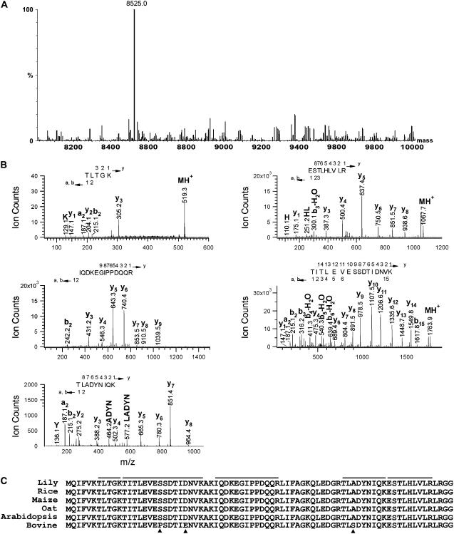
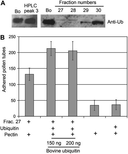
Pollen tube adhesion and guidance on extracellular matrices within the pistil are essential processes that convey the pollen tube cell and the sperm cells to the ovule. In this study, we purified an additional molecule from the pistil that enhances pollen tube adhesion when combined with the SCA (stigma/stylar cysteine-rich adhesin)/pectin matrix in our in vitro assay. The enhancer of adhesion was identified as free ubiquitin (Ub). This was confirmed by use of bovine Ub as a substitute for lily (Lilium longiflorum Thunb.) stigma Ub. To study the interaction of SCA and Ub with the lily pollen tube, we labeled both proteins with biotin. We observed uptake of biotin-labeled SCA and Ub into the pollen tube cells in vitro using confocal microscopy. For SCA, a strong signal occurred first at the tip of the pollen tube, suggestive of an endocytosis event, and then progressively throughout the tube cytoplasm. SCA was also localized inside the in vivo pollen tube using immunogold electron microscopy and found to be present in endosomes, multivesicular bodies, and vacuoles, all known to be endocytic compartments. It was also confirmed that SCA is endocytosed in the in vitro adhesion assay. Internalization of SCA was increased in pollen tubes treated with exogenous Ub compared to those without Ub, suggesting that Ub may facilitate SCA endocytosis. These results show that Ub can act as an enhancer of pollen tube adhesion in vitro and that it is taken up into the pollen tube as is SCA. The Ub machinery may play a role in pollen tube adhesion and guidance in lily.


Figures

Figure 1.
Figure 1.
SP purification. A, Total PBS extracts (T) were precipitated with various concentrations (50%, 60%, 70%, and 80%) of methanol and then separated as two fractions, pellet and supernatant. B, Among them, the 70% methanol supernatant was subjected to the CM-Sephadex C-25 cation exchange column and eluted stepwise with 0.1 and 0.25 m NaCl in 20 mm MES buffer. C, To further separate elutes 1 and 2 in 0.25 m NaCl (B), they were fractionated on Bio-Gel P-10 gel (Bio-Rad). Fraction size in B and C: 20 mL and 1.5 mL, respectively. Five micrograms of proteins was loaded into the SDS-PAGE gel. Western-blot analysis of gels using purified polyclonal antibodies raised from HPLC-purified SCA (Anti-SCA) and Escherichia coli-expressed chemocyanin (Anti-Chemo) were performed. S, Methanol supernatant fraction. UB, Unbound proteins. SP, Starting material as a positive control. Molecular mass standards (in kD) are shown on the left. Blot (C) was stained with Ponceau S. The secondary antibody was an affinity-purified goat anti-rabbit IgG alkaline phosphatase and blots were detected with NBT/BCIP.
Figure 2.
Figure 2.
Fraction 30 contains SCA and the unknown enhancer of pollen tube adhesion activity. Adhesion assays were done using fractions 27 to 30. A, For each assay, 5 μg of fractionated SCA and 25 μg of pectin were used. Inserted box in A indicates a loading control. Proteins were separated on a 15% acrylamide gel and stained with Coomassie Brilliant Blue. Bars indicate the sd from the mean for three replicates. B, ESI-MS spectra of fractions 27 and 30. Peaks at m/z 9,369.4 and 9,369.6 correspond to the SCA1 molecular mass, which predominated in both fractions 27 and 30.
Figure 3.
Figure 3.
Further separation of fraction 30 reveals the enhancer of adhesion as peak 3. A and B, HPLC profile of fraction 30 (A), SDS gel stained with Coomassie Brilliant Blue, and western blot (WB; B) of peak 2 against anti-SCA antibody. The secondary antibody was used with an affinity-purified goat anti-rabbit IgG alkaline phosphatase and blots were detected with NBT/BCIP. B, Proteins were separated on a 15% acrylamide gel and stained with Coomassie Brilliant Blue. Lane 1 is fraction 30 prior to HPLC (5 μg), lane 2 is HPLC-purified SCA (peak 2 in A, 5 μg), and lane 3 is a mixture of HPLC-purified SCA (peak 2 in A, 5 μg) and peak 3 (50 ng). Molecular mass standards (in kD) are shown at left. C, Adhesion assays. For each assay, 5 μg of HPLC-purified SCA and 25 μg of pectin were used. Bars indicate the sd from the mean for three replicates. D shows adhesion assay using 25 μg of pectin and, for the SCA source, fraction 30, peak 2 alone, and peaks 2 and 3 combined. Assays were stained with Coomassie Brilliant Blue to detect adhered pollen tubes.
Figure 4.
Figure 4.
Peak 3 is Ub. A, Spectrum of ESI-MS of HPLC-purified peak 3. B, The major peak was eluted at m/z 8,525 D. Shown is nano LC-MS/MS sequence analysis of trypsin-digested peptides from peak 3. The analyzed peptide sequences (TITLEVESSDTIDNVK, ESTLHLVLR, TLADYNIQK, IQDKEGIPPDQQR, and TLTGK) correspond to Ub. C, This is indicated along with the b (N-terminal) and y (C-terminal) fragment ions. Full-length amino acid sequence of lily Ub was compared with plant and bovine Ub. The peptides identified by nano LC-MS/MS in Ub are underlined. Arrowheads indicate the different amino acids compared to bovine Ub.
Figure 5.
Figure 5.
Bovine Ub can substitute for peak 3 in the pollen tube adhesion assay. A, Western-blot analysis of HPLC-purified peak 3 and fractions 27 to 30 was carried out using anti-human Ub. Bovine Ub (Bo; 200 ng) was used as a positive control. Ten micrograms of protein from each fraction was loaded into the SDS-PAGE gel. After treatment with primary antibody (dilution 1/200), the blot was incubated with an affinity-purified goat anti-rabbit IgG horseradish peroxidase-conjugated secondary antibody (Bio-Rad), which was detected by the enhanced chemiluminescence system. B, Adhesion assays were conducted with lily pollen tubes on a pectin matrix coated with SP fraction 27 (SCA) and bovine Ub. For each assay, 5 μg of fraction 27 (SCA) and 25 μg of pectin were used. Bovine Ub (150 ng and 200 ng) was also used. As negative controls, pectin alone and a pectin/Ub mixture without SCA were used. Bars indicate the sd from the mean for three replicates.
Figure 6.
Figure 6.
SCA binds to the pollen tube tip and is endocytosed. A, Labeled (LSCA) and unlabeled (SCA) SCA were run in the SDS-PAGE gels. Left and right gels are reduced and nonreduced gels, respectively. Blots were stained with Ponceau S. The signal was detected with a streptavidin conjugated with horseradish peroxidase. Molecular mass standards (in kD) are shown at left in lane 1. B, Comparison of activities of LSCA and unlabeled SCA in the adhesion assay. For this assay, 10 μg of SCA or LSCA and 50 μg of pectin were used to make the matrix. Bars indicate the sd from the mean for three replicates. C, Localization of LSCA using Sulfo-SBED. Pollen tubes were treated with 100 μg/mL LSCA, Sulfo-SBED alone, and LBSA (100 μg/mL). The signal was detected with streptavidin conjugated with FITC (1/200). D, Immunolocalization of SCA in adhered pollen tubes using purified SCA antibody. Lily pollen tube adhesion assay was performed on cellulose sheets coated with pectin and SCA. Adhered and in vitro-grown pollen tubes were fixed, sectioned, and analyzed with antibodies against SCA (dilution 1/50). FITC-conjugated secondary antibody (dilution 1/100) was used to detect signals. Pollen tubes were examined under a confocal microscope (TCS SP2; Leica) at 488 nm excitation. Bars represent 10 μm.
Figure 7.
Figure 7.
Immunogold localization of SCA in the TTE of the style and in vivo pollen tubes. Thin sections were prepared from chemically fixed specimens and immunostained with affinity-purified antibodies against HPLC-purified SCA (dilution 1/5). A (secondary antibody, control) and B (anti-SCA), Localization of SCA on lily TTE. C (secondary antibody, control) to H, Localization of SCA in in vivo-grown pollen tubes (D to J: anti-SCA). Arrows indicate internal vesicles. cyt, Cytoplasm of TTE; C, stylar canal; g, Golgi; mvb, multivesicular body; OW, outer wall of TTE; V, vacuole. Bars represent 200 nm.
Figure 8.
Figure 8.
Uptake of LUb into the pollen tube. The N-terminal LUb was purchased from BostonBiochem. Complied confocal images are displayed. Pollen tubes were harvested at 10 min after treatment of LUb (10 μg/mL). As a negative control, Sulfo-SBED alone (Biotin alone) was used. The signal was detected with streptavidin conjugated with FITC. The pollen tubes were examined under a confocal microscope (TCS SP2; Leica) at 488 nm excitation. Bars represent 10 μm.
Figure 9.
Figure 9.
Immunogold localization of Ub in the stylar TTE and in vivo pollen tubes. Thin sections were prepared from chemically fixed specimens and immunostained with anti-Ub from Santa Cruz Biotechnology (dilution 1/10). A, Localization of Ub on lily TTE. B, Localization of Ub in in vivo-grown pollen tubes. cyt, Cytoplasm of TTE; C, stylar canal; OW, outer wall of TTE; V, vacuole. Bars represent 200 nm.
Figure 10.
Figure 10.
Western-blot analysis of SCA in in vitro-grown pollen tubes exposed to Ub and SCA in the medium. Pollen tubes grown in vitro were harvested at 30 min after treatment of SCA and bovine Ub (bUb). Left, The first lane is total pollen tube proteins without any treatment of SCA or BUb (C). The second lane is 10 μg/mL SP fraction without lily Ub (Fraction). The third lane is 10 μg/mL SP fraction plus 1 μg/mL bovine Ub (Fraction + bUb). Right, The first lane is 10 μg/mL HPLC-purified SCA (HPLC SCA). The second lane is HPLC SCA plus 1 μg/mL bovine Ub (HPLC SCA + bUb). The third lane is fraction 30 (SCA + peak 3) as a positive control. Pollen tubes were washed to remove nonspecific SCA from the pollen tube wall. Proteins were extracted from the pollen tubes with SDS-PAGE buffer. Ten micrograms of total protein extracted was loaded in each lane. Blot (Anti-SCA) was immunostained with affinity-purified antibodies against HPLC-purified SCA (dilution 1/5). P, Ponceau S.
Figure 11.
Figure 11.
A model for endocytosis of SCA in pollen tube adhesion. A, Ub-dependent internalization of GPCR in Saccharomyces cerevisiae. Interaction of mating factor with a GPCR activates a signal transduction pathway and subsequently stimulates endocytosis of the receptor-ligand complex by ubiquitination. Modification of the receptor with monoubiquitination in the cytoplasmic side of the GPCR promotes rapid internalization, followed by degradation in the vacuole. B, SCA binds to its putative receptor (SCAR), which may be present at the membrane in the pollen tube tip. This binding would activate the signals for pollen tube adhesion, which are currently unknown. Subsequently, SCA is endocytosed through the endocytic route (early endosome, MVB, and vacuole). PM, Plasma membrane. The yeast model is from Hicke (1999) with some modification.

Similar articles

Cited by

References

    1. Bahaji A, Cornejo MJ, Ortiz-Zapater E, Contreras I, Aniento F (2001) Uptake of endocytic markers by rice cells: variations related to the growth phase. Eur J Cell Biol 80: 178–186 - PubMed
    1. Beers EP, Moreno TM, Callis J (1992) Subcellular localization of ubiquitin and ubiquitinated proteins in Arabidopsis thaliana. J Biochem (Tokyo) 267: 15432–15439 - PubMed
    1. Blackbourn HD, Jackson AP (1996) Plant clathrin heavy chain: sequence analysis and restricted localisation in growing pollen tubes. J Cell Sci 109: 777–787 - PubMed
    1. Blein JP, Coutos-Thevenot P, Marion D, Ponchet M (2002) From elicitins to lipid-transfer proteins: a new insight in cell signalling involved in plant defence mechanisms. Trends Plant Sci 7: 293–296 - PubMed
    1. Buhot N, Douliez JP, Jacquemard A, Marion D, Tran V, Maume BF, Milat ML, Ponchet M, Mikes V, Kader JC, et al (2001) A lipid transfer protein binds to a receptor involved in the control of plant defence responses. FEBS Lett 509: 27–30 - PubMed

Publication types

MeSH terms

LinkOut - more resources