. 2006;34(18):5133-44.
doi: 10.1093/nar/gkl610. Epub 2006 Sep 22.

NMR solution structure of the major G-quadruplex structure formed in the human BCL2 promoter region

Affiliations

NMR solution structure of the major G-quadruplex structure formed in the human BCL2 promoter region

Jixun Dai et al. Nucleic Acids Res. 2006.

Abstract

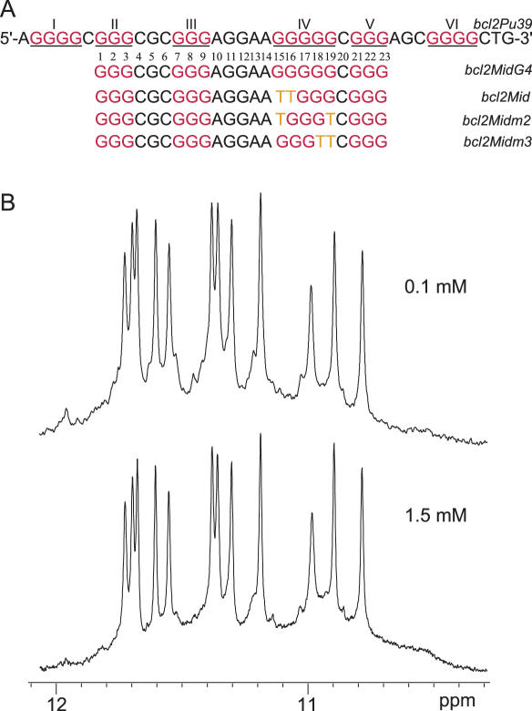
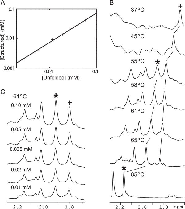
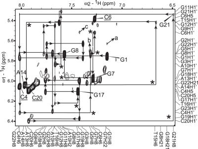
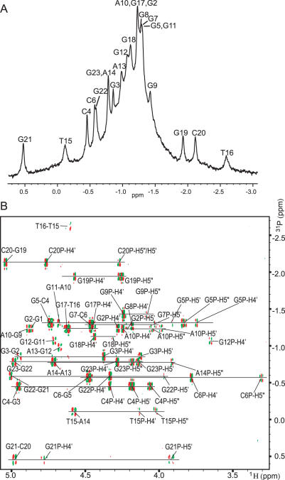
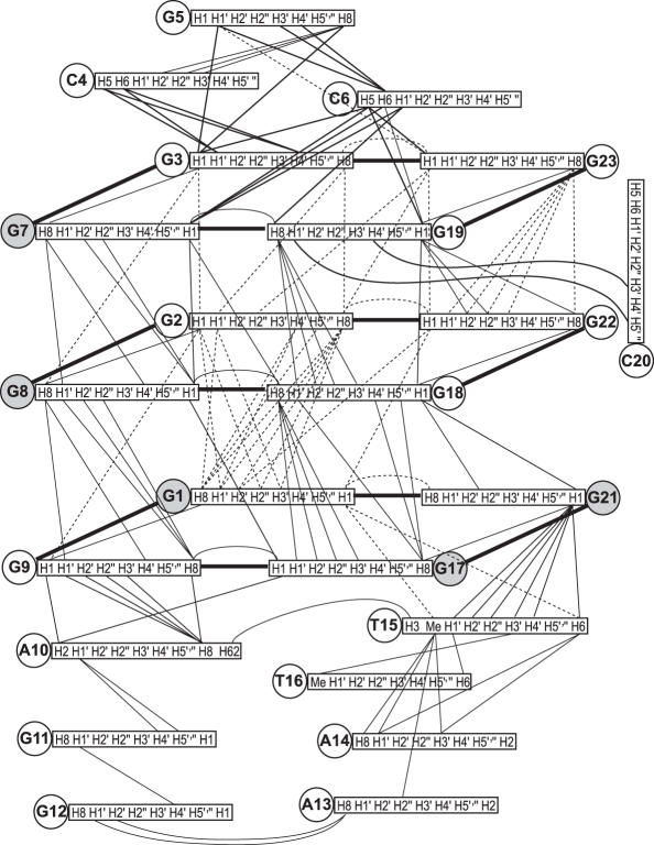
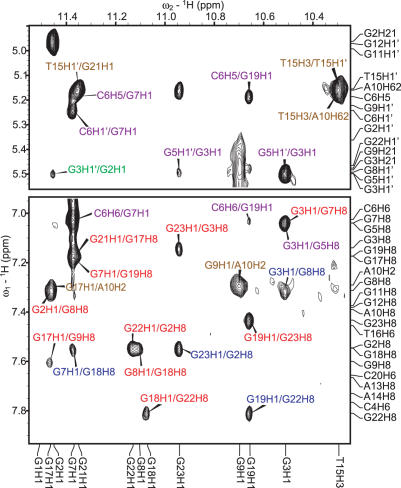
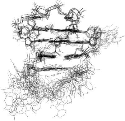
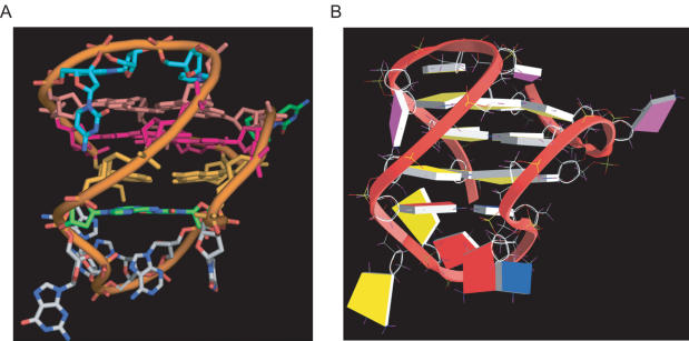
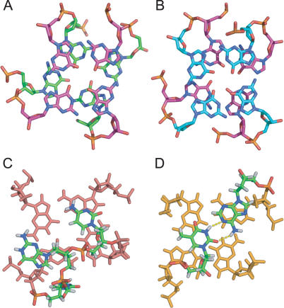
BCL2 protein functions as an inhibitor of cell apoptosis and has been found to be aberrantly expressed in a wide range of human diseases. A highly GC-rich region upstream of the P1 promoter plays an important role in the transcriptional regulation of BCL2. Here we report the NMR solution structure of the major intramolecular G-quadruplex formed on the G-rich strand of this region in K+ solution. This well-defined mixed parallel/antiparallel-stranded G-quadruplex structure contains three G-tetrads of mixed G-arrangements, which are connected with two lateral loops and one side loop, and four grooves of different widths. The three loops interact with the core G-tetrads in a specific way that defines and stabilizes the overall G-quadruplex structure. The loop conformations are in accord with the experimental mutation and footprinting data. The first 3-nt loop adopts a lateral loop conformation and appears to determine the overall folding of the BCL2 G-quadruplex. The third 1-nt double-chain-reversal loop defines another example of a stable parallel-stranded structural motif using the G3NG3 sequence. Significantly, the distinct major BCL2 promoter G-quadruplex structure suggests that it can be specifically involved in gene modulation and can be an attractive target for pathway-specific drug design.


Figures

Figure 1
Figure 1
(A) The promoter sequence of the BCL2 gene and its modifications. Bcl2Pu39 is the wild-type BCL2 39mer sequence; bcl2MidG4 is the BCL2 23mer sequence containing the middle four consecutive G-runs, which forms the most stable G-quadruplex structure; bcl2Mid is the mutant 23mer with G-to-T mutations at positions 15 and 16, bcl2Midm2 is the mutant 23mer with G-to-T mutations at positions 15 and 19, and bcl2Midm3 is the mutant 23mer with G-to-T mutations at positions 18 and 19. The six G-runs are underlined and numbered using Roman numerals; the BCL2 23mer is numbered using Arabic numerals. (B) The imino regions of 1D 1H NMR spectra of bcl2Mid samples at 0.1 mM (upper) and 1.5 mM (lower) strand concentrations. Conditions: 25°C, 20 mM K-phosphate, 40 mM KCl, pH 7.0.
Figure 2
Figure 2
(A) Determination of stoichiometry by NMR titration for bclMid in K+ solution. The slope of the fitted line is 0.99, meaning that the quadruplex structure existing in solution is unimolecular. (B) The variable-temperature study of bcl2Mid by NMR. The peak intensities of two resolved peaks at 61°C (the one belonging to the melted form of bcl2Mid is labeled with asterisk and the one belonging to the folded forms is labeled with cross) were used for the calculation. (C) The extended region of 1D 1H NMR spectra of bcl2Mid at various concentrations showing the two peaks from the folded and the melted forms. Conditions: 20 mM K-phosphate, 40 mM KCl, pH 7.0.
Figure 3
Figure 3
(A) (Left) Schematic drawing of the folding topology of bcl2Mid. Red, guanine (anti); light red, guanine (syn); green, adenine; yellow, thymine; blue, cytosine. (Right) A G-tetrad with H1–H1 and H1–H8 connectivity pattern detectable in NOESY experiments. (B) Imino and aromatic regions of the 1D 1H NMR spectrum of bcl2Mid. The imino and aromatic protons are assigned over the resonances. Conditions: 25°C, 20 mM K-phosphate, 40 mM KCl, pH 7.0, 1.5 mM DNA.
Figure 4
Figure 4
The expanded H8/H6–H1′ region with assignments of the non-exchangeable 2D-NOESY spectrum of bcl2Mid.The sequential assignment pathway is shown. Missing connectivities are labeled with asterisks. The H8–H1′ NOEs of the nucleotides with syn configuration are labeled by residue names, while the H5–H6 NOEs of cytosines are also labeled for reference. The H8–H1′ NOE crosspeak of G21 has a large integration value; however, the H8 and H1′ of G21 are much broader than those of other guanines and give rise to a much broader H8–H1′ NOE crosspeak. The characteristic G(i)H8/G(i+1)H1′ NOEs for the syn G(i)s are labeled as a–b for G1–G2 and G7–G8. The NOE between C6H1′ and G19H8 is labeled as c. Conditions: 25°C, 20 mM K-phosphate, 40 mM KCl, pH 7.0, 1.5 mM DNA.
Figure 5
Figure 5
(A) 1D proton-decoupled 31P NMR spectrum of bcl2Mid with phosphorus assignments. (B) 2D heteronuclear 31P–1H Correlation Spectroscopy (COSY) of bcl2Mid with peak assignments. The NnP-N(n-1)H3′ crosspeaks are labeled as Nn-N(n-1). The spectra were referenced to external H3PO4. Conditions: 25°C, 20 mM K-phosphate, 40 mM KCl, pH 7.0, 1.5 mM DNA.
Figure 6
Figure 6
Schematic diagram of inter-residue NOE connectivities of bcl2Mid. The guanines in syn configuration are represented using gray circles. The NOE connectivities clearly define the quadruplex conformation and provide distance restraints for structure calculation.
Figure 7
Figure 7
The expanded H1–H8/H6 and H1–H1′ regions with assignments of the exchangeable proton 2D-NOESY spectrum of bcl2Mid. For NOEs involved in G-tetrads: intra-tetrad NOEs are labeled in red, sequential NOEs are labeled in green, and inter-tetrad NOEs are labeled in blue. NOEs involved in the first C4–G5–C6 lateral loop region are labeled in purple, and NOEs involved in the second A10–G11–G12–A13–A14–T15–T16 lateral loop region are labeled in brown. Conditions: 1°C, 20 mM K-phosphate, 40 mM KCl, pH 7.0, 1.5 mM DNA.
Figure 8
Figure 8
The superimposed 10 lowest energy structures of the bcl2Mid G-quadruplex by NOE-restrained structure refinement.
Figure 9
Figure 9
A representative model of the NMR-refined bcl2Mid G-quadruplex structure from two different views. (A) is prepared using PyMOL. (B) is prepared using GRASP (57) (guanine, yellow; adenine, red; thymine, blue; and cytosine, magenta).
Figure 10
Figure 10
Stacking interactions between (A) the middle (magenta) and bottom (green) G-tetrads, which have reversed anti:syn:anti:anti and syn:anti:syn:syn arrangements, and (B) the top (cyan) and middle (magenta) G-tetrads, which have the same anti:syn:anti:anti arrangements; and stacking interactions between (C) the first lateral loop C4–G5–C6 (cyan) and the top G-tetrad (pink), and (D) the bottom G-tetrad (orange) and the A10:T15 bp (green) from the second lateral loop, A10–G11–G12–A13–A14–T15–T16. Figures are prepared using PyMOL.

References

    1. Adams J.M., Cory S. The Bcl-2 protein family: arbiters of cell survival. Science. 1998;281:1322–1326. - PubMed
    1. Chao D.T., Korsmeyer S.J. BCL-2 family: regulators of cell death. Annu. Rev. Immunol. 1998;16:395–419. - PubMed
    1. Cleary M.L., Smith S.D., Sklar J. Cloning and structural analysis of cDNAs for bcl-2 and a hybrid bcl-2/immunoglobulin transcript resulting from the t(14;18) translocation. Cell. 1986;47:19–28. - PubMed
    1. Akagi T., Kondo E., Yoshino T. Expression of Bcl-2 protein and Bcl-2 mRNA in normal and neoplastic lymphoid tissues. Leuk. Lymphoma. 1994;13:81–87. - PubMed
    1. Joensuu H., Pylkkanen L., Toikkanen S. Bcl-2 protein expression and long-term survival in breast cancer. Am. J. Pathol. 1994;145:1191–1198. - PMC - PubMed

Publication types