Review
. 2006 Oct;28(10):983-93.
doi: 10.1002/bies.20477.

Rho GTPase activity zones and transient contractile arrays

Affiliations
Review

Rho GTPase activity zones and transient contractile arrays

William M Bement et al. Bioessays. 2006 Oct.

Abstract

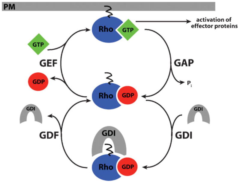
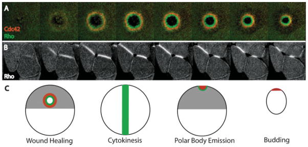
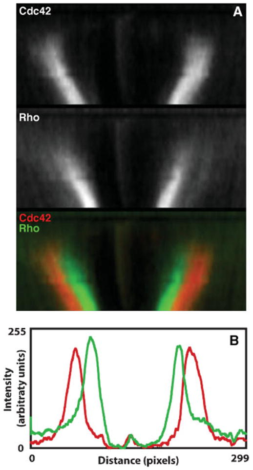
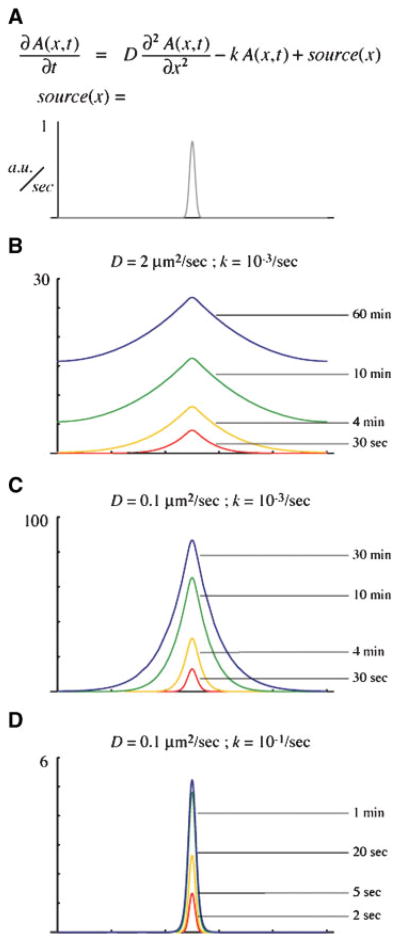
The Rho GTPases-Rho, Rac and Cdc42-act as molecular switches, cycling between an active GTP-bound state and an inactive GDP-bound state, to regulate the actin cytoskeleton. It has recently become apparent that the Rho GTPases can be activated in subcellular zones that appear semi-stable, yet are dynamically maintained. These Rho GTPase activity zones are associated with a variety of fundamental biological processes including symmetric and asymmetric cytokinesis and cellular wound repair. Here we review the basic features of Rho GTPase activity zones, suggest that these zones represent a fundamental signaling mechanism, and discuss the implications of zone properties from the perspective of both their function and how they are likely to be controlled.


Figures

Figure 1
Figure 1
The Rho GTPase cycle. Rho GTPases cycle between an active, GTP-bound state and an inactive, GDP-bound state. Their activity is regulated by four factors: (1) Guanine nucleotide exchange factors (GEFs) activate Rho GTPases by promoting the exchange of GDP for GTP, (2) GTPase activating proteins (GAPs) promote GTP hydrolysis, (3) guanine nucleotide dissociation inhibitors (GDIs) stabilize the GDP-bound state and mask the lipid moeity, maintaining Rho GTPases in an inactive state in the cytoplasm, and (4) GDI displacement factors (GDFs) disrupt GDI-GTPase binding, facilitating GTPase activation. Only when Rho is in its active, GTP-bound conformation at the plasma membrane (PM) can it interact with downstream effector proteins to modulate the cytoskeleton.
Figure 2
Figure 2
Rho activity zones regulate transient contractile arrays in diverse cellular contexts. A:Active Rho (green) and active Cdc42 (red) segregate into discrete zones during wound healing. Active Rho is concentrated in a ring that is circumscribed by a ring of active Cdc42. B: Rho activity zones predict the site of cleavage furrow formation during cytokinesis. The Rho activity zones form a stripe-like zone that remains tightly focused and moves inward in concert with the ingressing cleavage furrow. C: Rho GTPase activity zones are employed in diverse contexts including wound healing, cytokinesis, polar body emission and budding. Although the overall geometric organization may vary, the Rho activity zones share many common features (see text for details).
Figure 3
Figure 3
Rho activity zones are spatially segregated and tightly focused. Two common features of Rho activity zones are their sharp boundaries and spatial segregation. These features are illustrated here for the case of wound healing (see also Ref. for an example from cytokinesis). A: Kymographs of active Cdc42 (red) and active Rho (green) during wound healing. Time is in the vertical direction with regions from successive time points aligned from top to bottom for each panel. The zones remain spatially segregated with active Rho localized on the inside, Cdc42 on the outside, and little overlap between the two. B: Line scans of the intensities of active Rho (green) and active Cdc42 (red). Notice that the peaks are tightly focused, with sharp boundaries between regions containing active or inactive GTPases.
Figure 4
Figure 4
Numerical solutions of a simple local source/global sink reaction–diffusion equation. A: The partial differential equation relates the rate of change of the concentration of some diffusible species “A”, at each point in space and time, to the spatial gradient (first term) and to the local concentration (second term). “D” is the diffusion coefficient, and “k” is the first-order decay rate. The source is a narrow Gaussian centered in the middle of the domain which creates “A” at a peak rate of ~1 arbitrary unit per second. The entire domain is 100 microns wide, and the edges are constrained to have the same level of “A” (periodic boundary conditions). B–D: Numerical solutions, computed using Mathematica, in which the concentration of “A” is plotted in arbitrary units across the entire domain. Four successive timepoints are shown for each of three conditions. B: With a diffusion coefficient similar to myristoylated yellow fluorescent protein and a decay rate similar to the intrinsic rate for Rho GTP hydrolysis, within seconds “A” has already appeared far from the source, and the domain slowly fills up with “A”. C: With a much smaller diffusion coefficient, the profile of “A” is still much wider than the source and also requires tens of minutes to reach steady state. D: The combination of slow diffusion and rapid turnover leads to a narrow zone, which reaches steady state quickly.
Figure 5
Figure 5
The GTPase flux model. Rho activity zones are maintained by a balance of local GTPase activation (by GEFs) with local GTPase inactivation (by GAPs). This would result in the constant flux of Rho through the GTPase cycle, allowing cells to maintain tightly focused, dynamic Rho activity zones. In this way, any active Rho that does not immediately interact with an effector protein will be switched back off.

References

    1. Kishi K, Sasaki T, Kuroda S, Itoh T, Takai Y. Regulation of cytoplasmic division of Xenopus embryo by rho p21 and its inhibitory GDP/GTP exchange protein (rho GDI) J Cell Biol. 1993;120:1187–1195. - PMC - PubMed
    1. Brock J, Midwinter K, Lewis J, Martin P. Healing of incisional wounds in the embryonic chick wing bud: characterization of the actin purse-string and demonstration of a requirement for Rho activation. J Cell Biol. 1996;135:1097–1107. - PMC - PubMed
    1. Benink HA, Bement WM. Concentric zones of active RhoA and Cdc42 around single cell wounds. J Cell Biol. 2005;168:429–439. - PMC - PubMed
    1. Wood W, Jacinto A, Grose R, Woolner S, Gale J, et al. Wound healing recapitulates morphogenesis in Drosophila embryos. Nat Cell Biol. 2002;4:907–912. - PubMed
    1. Etienne-Manneville S, Hall A. Rho GTPases in cell biology. Nature. 2002;420:629–635. - PubMed