. 2006 Nov 20;499(3):506-18.
doi: 10.1002/cne.21113.

Distinct and gradient distributions of connexin26 and connexin30 in the cochlear sensory epithelium of guinea pigs

Affiliations

Distinct and gradient distributions of connexin26 and connexin30 in the cochlear sensory epithelium of guinea pigs

Hong-Bo Zhao et al. J Comp Neurol. .

Abstract

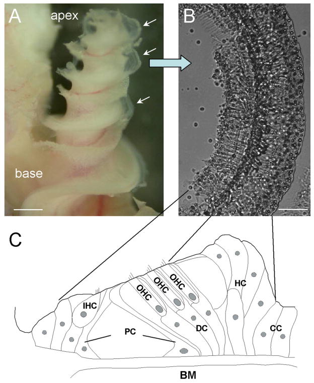
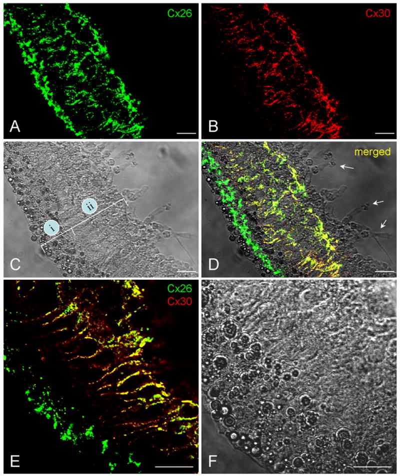
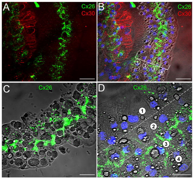
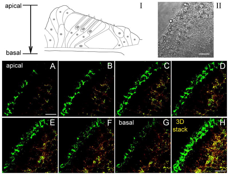
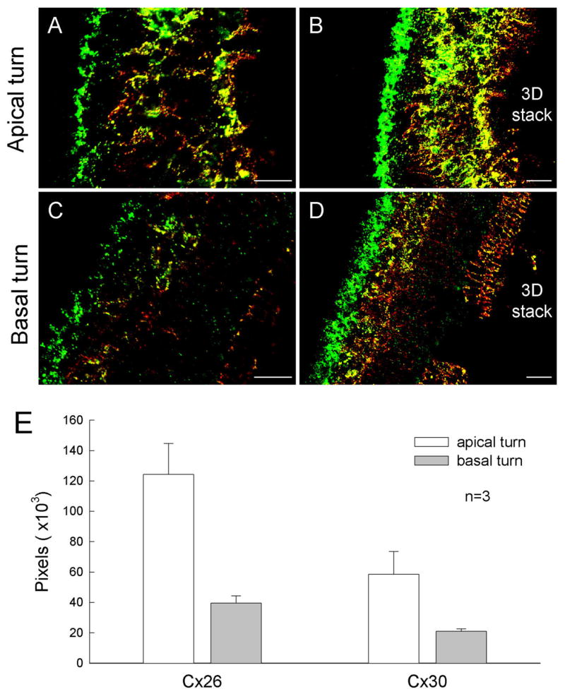
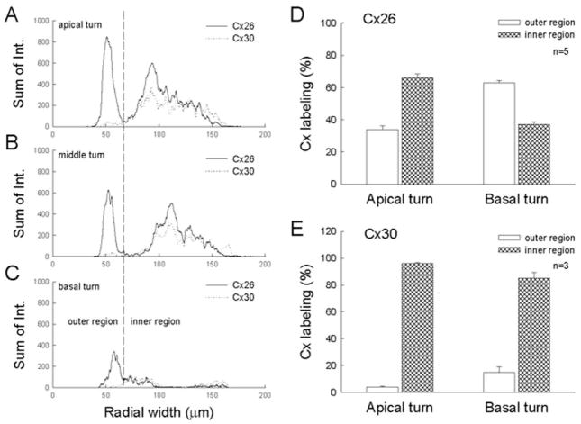
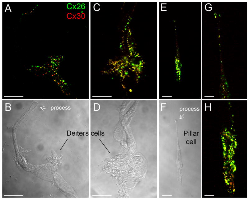
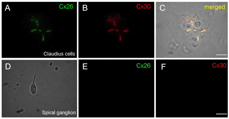
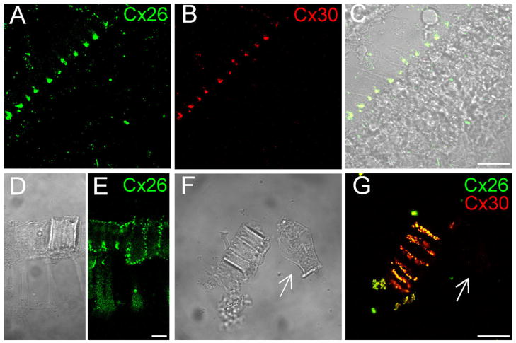
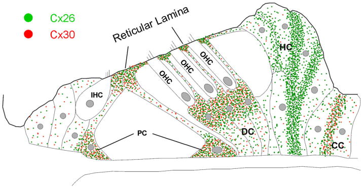
Connexin26 (Cx26) and Cx30 are predominant isoforms of gap junction channels in the cochlea and play a critical role in hearing. In this study, the cellular distributions of Cx26 and Cx30 in the cochlear sensory epithelium of guinea pigs were examined by immunofluorescent staining and confocal microscopy in whole mounts of the cochlear sensory epithelium and dissociated cell preparations. The expression of Cx26 and Cx30 demonstrated a longitudinal gradient distribution in the epithelium and was reduced threefold from the cochlear apex to base. The reduction was more pronounced in the Deiters cells and pillar cells than in the Hensen cells. Cx26 was expressed in all types of supporting cells, but little Cx30 labeling was seen in the Hensen cells. Cx26 expression in the Hensen cells was concentrated mainly in the second and third rows, forming a distinct band along the sensory epithelium at its outer region. In the dissociated Deiters cells and pillar cells, Cx30 showed dense labeling at the cell bodies and processes in the reticular lamina. Cx26 labeling largely overlapped that of Cx30 in these regions. Cx26 and Cx30 were also coexpressed in the gap junctional plaques between Claudius cells. Neither Cx26 nor Cx30 labeling was seen in the hair cells and spiral ganglion neurons. These observations demonstrate that Cx26 and Cx30 have a longitudinal gradient distribution and distinct cellular expression in the auditory sensory epithelium. This further supports our previous reports that Cx26 and Cx30 can solely and concertedly perform different functions in the cochlea.


Figures

Fig. 1
Fig. 1
Structure of the cochlea and the organ of Corti. A: Micrograph of the guinea pig cochlea after removal of the bone and stria vascularis. The auditory sensory epithelium is visible and is indicated by arrows. B: The isolated sensory epithelium. C: Diagram of the organ of Corti in a cross-section of the epithelium. IHC, inner hair cell; OHC, outer hair cell; PC, pillar cell; DC, Deiters cell; HC, Hensen cell; CC, Claudius cell; BM, basilar membrane. Scale bars = 0.5 mm in A; 100 μm in B.
Fig. 2
Fig. 2
Double-immunofluorescent staining of the cochlear sensory epithelium for Cx26 and Cx30 in whole-mount preparation. Green and red represent immunofluorescent staining for Cx26 and Cx30, respectively. A,B: The confocal images of immunofluorescent staining for Cx26 and Cx30. C: Nomarski image. Lines indicate the outer epithelial region (i) and inner epithelial region (ii). D: Merged image of immunofluorescent staining with Nomarski image. Arrows indicate outer hair cells that have no immunofluorescent labeling. E,F: High-magnification images of the epithelium stained for Cx26 and Cx30. F is a Nomarski image in the same field. Scale bars = 25 μm in A–D; 20 μm in E,F.
Fig. 3
Fig. 3
Connexin expression in the Hensen cells at the outer epithelial region. A: Immunofluorescent image of double staining of the epithelium for Cx26 and Cx30. B: Superimposed image of fluorescent staining with Nomarski image. Blue represents cell nuclei stained by DAPI. Lines and circled numbers indicate four rows of Hensen cells. C,D: High-magnification images in the Hensen cell region. Cx26 labeling is located mainly at the second and third rows of Hensen cells. Scale bars = 40 μm in A–C; 20 μm in D.
Fig. 4
Fig. 4
Immunofluorescent staining for Cx26 and Cx30 in the cochlear sensory epithelium along the Z-axis. Serial confocal sections were scanned by a 2.2-μm step from the apical epithelial surface down to its basal bottom (I). Green and red colors represent Cx26 and Cx30 staining, respectively. II is a Nomarski image. Scale bars = 10 μm in I,II; 10 μm in A,H (apply to A–H).
Fig. 5
Fig. 5
Different expressions of Cx26 and Cx30 in the epithelia from the apical and basal turns. A: High-magnification image of immunofluorescent staining of the epithelium from the apical turn. B: Low-magnification image stacked from all scanning sections along the Z-axis. C,D: Single confocal section and 3D-stack immunofluorescent image of the epithelium from the basal turn. E: Quantitative analysis of Cx26 and Cx30 expressions in the apical and basal turns. The epithelia were isolated from the apical and basal turns in the same cochlea and stained at the same time. The pixels of positive staining for Cx26 and Cx30 were separately counted. Data from three animals were averaged. Error bars represent SE. Scale bars = 10 μm in A,C; 25 μm in B,D.
Fig. 6
Fig. 6
Comparison of Cx26 and Cx30 expressions at the inner and outer epithelial regions in the apical and basal turns. A–C: Intensities of Cx26 and Cx30 staining were measured along the radial direction of the epithelia from the apical, middle, and basal turns in the same cochlea. The epithelium was serially scanned along the Z-axis, and the intensities of staining for Cx26 and Cx30 were separately measured in each section, then summed. D,E: Percentages of Cx26 and Cx30 distributions at the inner and outer epithelial regions in the apical and basal turns. Error bars represent SE.
Fig. 7
Fig. 7
A–H: Double-immunofluorescent staining of Deiters cells and a pillar cell for Cx26 and Cx30 in dissociated cell preparation. G and H are high-magnification images of E. Scale bar = 20 μm in A–D; 25μm in E,F; 10 μm in G,H.
Fig. 8
Fig. 8
A–C: Double-immunofluorescent staining of Claudius cells for Cx26 and Cx30. C is a merged image showing coexpression of Cx26 and Cx30 in the same gap junctional plaques between cells. D–F: Immunofluorescent images of a spiral ganglion cell staining for Cx26 and Cx30. There is no fluorescent labeling in the cochlear spiral ganglion neuron. Scale bars = 15 μm in C,F (apply to A–F).
Fig. 9
Fig. 9
Connexin expression in the reticular lamina. A–C: Confocal images scanned on the apical surface of the reticular lamina. D,E: Cx26 expression in a piece of the dissociated reticular lamina. The reticular lamina shows a thick layer composed of the process heads of pillar cells and Deiters cells. Cx26 labeling is distributed on the outer and inner surfaces of the layer and along the junction lines between the process heads. F,G: Double-immunofluorescent staining for Cx26 and Cx30 in a piece of the dissociated reticular lamina. Arrows indicate an outer hair cell that has no fluorescence labeling in the same field. Scale bars = 20 μm in C,G (apply to A–C,F,G); 10 in E (applies to D,E).
Fig. 10
Fig. 10
Schematic diagram of the cellular and subcellular distributions of Cx26 and Cx30 in the cochlear sensory epithelium. Green and red spots represent Cx26 and Cx30 expression, respectively. IHC, inner hair cell; OHC, outer hair cell; PC, pillar cell; DC, Deiters cell; HC, Hensen cell; CC, Claudius cell.

References

    1. Beltramello M, Bicego M, Piazza V, Ciubotaru CD, Mammano F, D’Andrea P. Permeability and gating properties of human connexins 26 and 30 expressed in HeLa cells. Biochem Biophys Res Commun. 2003;305:1024–1033. - PubMed
    1. Bruzzone R, Veronesi V, Gomes D, Bicego M, Duval N, Marlin S, Petit C, D’Andrea P, White TW. Loss-of-function and residual channel activity of connexin26 mutations associated with nonsyndromic deafness. FEBS Lett. 2003;533:79–88. - PubMed
    1. Cheatham MA, Huynh KH, Gao J, Zuo J, Dallos P. Cochlear function in Prestin knockout mice. J Physiol. 2004;560:821– 830. - PMC - PubMed
    1. Choung YH, Moon SK, Park HJ. Functional study of GJB2 in hereditary hearing loss. Laryngoscope. 2002;112:1667–1671. - PubMed
    1. Cohen-Salmon M, Ott T, Michel V, Hardelin JP, Perfettini I, Eybalin M, Wu T, Marcus DC, Wangemann P, Willecke K, Petit C. Targeted ablation of connexin26 in the inner ear epithelial gap junction network causes hearing impairment and cell death. Curr Biol. 2002;12:1106–1111. - PMC - PubMed

Publication types

MeSH terms