

Intravenous administration of glial cell line-derived neurotrophic factor gene-modified human mesenchymal stem cells protects against injury in a cerebral ischemia model in the adult rat
- PMID: 16998918
- PMCID: PMC2605367
- DOI: 10.1002/jnr.21056
Intravenous administration of glial cell line-derived neurotrophic factor gene-modified human mesenchymal stem cells protects against injury in a cerebral ischemia model in the adult rat
Abstract
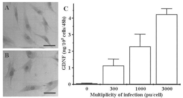
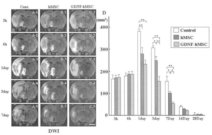
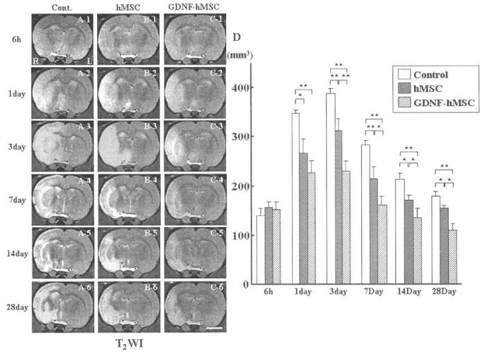
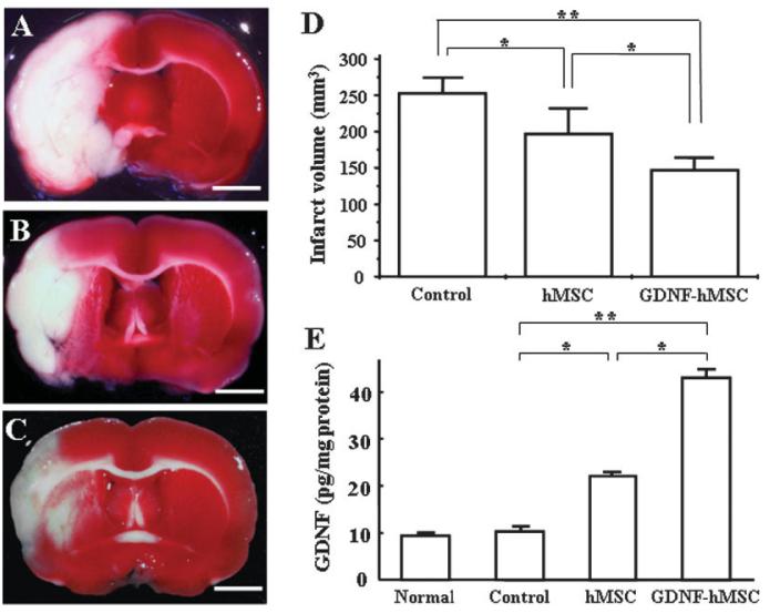
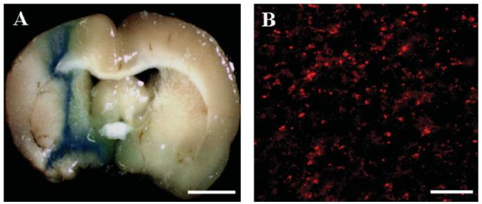
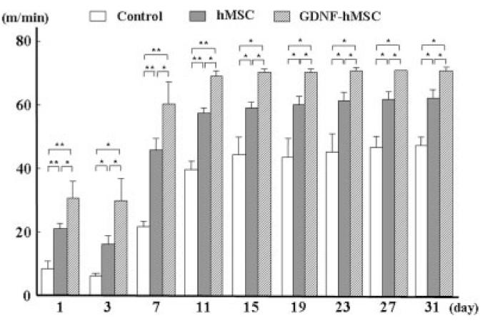
Intravenous administration of human mesenchymal stem cells (hMSCs) prepared from adult bone marrow has been reported to ameliorate functional deficits after cerebral artery occlusion in rats. Several hypotheses to account for these therapeutic effects have been suggested, and current thinking is that neuroprotection rather than neurogenesis is responsible. To enhance the therapeutic benefits of hMSCs potentially, we transfected hMSCs with the glial cell line-derived neurotrophic factor (GDNF) gene using a fiber-mutant F/RGD adenovirus vector and investigated whether GDNF gene-modified hMSCs (GDNF-hMSCs) could contribute to functional recovery in a rat permanent middle cerebral artery occlusion (MCAO) model. We induced MCAO by using intraluminal vascular occlusion, and GDNF-hMSCs were intravenously infused into the rats 3 hr later. MRI and behavioral analyses revealed that rats receiving GDNF-hMSCs or hMSCs exhibited increased recovery from ischemia compared with the control group, but the effect was greater in the GDNF-hMSC group. Thus, these results suggest that intravenous administration of hMSCs transfected with the GDNF gene using a fiber-mutant adenovirus vector may be useful in the cerebral ischemia and may represent a new strategy for the treatment of stroke.
Figures
Similar articles
-
Therapeutic benefits of angiogenetic gene-modified human mesenchymal stem cells after cerebral ischemia.Exp Neurol. 2009 Mar;216(1):47-55. doi: 10.1016/j.expneurol.2008.11.010. Epub 2008 Nov 27. Exp Neurol. 2009. PMID: 19094989
-
I.V. infusion of brain-derived neurotrophic factor gene-modified human mesenchymal stem cells protects against injury in a cerebral ischemia model in adult rat.Neuroscience. 2005;136(1):161-9. doi: 10.1016/j.neuroscience.2005.06.062. Epub 2005 Oct 17. Neuroscience. 2005. PMID: 16229956 Free PMC article.
-
Optimization of a therapeutic protocol for intravenous injection of human mesenchymal stem cells after cerebral ischemia in adult rats.Brain Res. 2008 Oct 21;1236:30-8. doi: 10.1016/j.brainres.2008.07.116. Epub 2008 Aug 9. Brain Res. 2008. PMID: 18722359 Free PMC article.
-
Recent Progress in Therapeutic Strategies for Ischemic Stroke.Cell Transplant. 2016;25(5):893-8. doi: 10.3727/096368916X690548. Epub 2016 Jan 18. Cell Transplant. 2016. PMID: 26786838 Review.
-
Adenoviral vector-mediated delivery of glial cell line-derived neurotrophic factor provides neuroprotection in the aged parkinsonian rat.Clin Exp Pharmacol Physiol. 2001 Nov;28(11):896-900. doi: 10.1046/j.1440-1681.2001.03544.x. Clin Exp Pharmacol Physiol. 2001. PMID: 11703392 Review.
Cited by
-
CdSe/ZnS Quantum Dots-Labeled Mesenchymal Stem Cells for Targeted Fluorescence Imaging of Pancreas Tissues and Therapy of Type 1 Diabetic Rats.Nanoscale Res Lett. 2015 Dec;10(1):959. doi: 10.1186/s11671-015-0959-3. Epub 2015 Jun 13. Nanoscale Res Lett. 2015. PMID: 26078050 Free PMC article.
-
Application of magnetic resonance imaging for monitoring stem cell transplantation for the treatment of cerebral ischemia.Neural Regen Res. 2012 Jun 5;7(16):1264-71. doi: 10.3969/j.issn.1673-5374.2012.16.009. Neural Regen Res. 2012. PMID: 25709625 Free PMC article.
-
Tracking mesenchymal stem cells using magnetic resonance imaging.Brain Circ. 2016 Jul-Sep;2(3):108-113. doi: 10.4103/2394-8108.192521. Epub 2016 Oct 18. Brain Circ. 2016. PMID: 30276283 Free PMC article. Review.
-
Current status of ischemic stroke treatment: From thrombolysis to potential regenerative medicine.Regen Ther. 2021 Oct 12;18:408-417. doi: 10.1016/j.reth.2021.09.009. eCollection 2021 Dec. Regen Ther. 2021. PMID: 34722837 Free PMC article. Review.
-
Intravascular stem cell transplantation for stroke.Transl Stroke Res. 2011 Sep;2(3):250-65. doi: 10.1007/s12975-011-0093-1. Epub 2011 Aug 4. Transl Stroke Res. 2011. PMID: 24323647
References
-
- Abe K, Hayashi T. Expression of the glial cell line-derived neurotrophic factor gene in rat brain after transient MCA occlusion. Brain Res. 1997;776:230–234. - PubMed
-
- Beck KD, Valverde J, Alexi T, Poulsen K, Moffat B, Vandlen RA, Rosenthal A, Hefti F. Mesencephalic dopaminergic neurons protected by GDNF from axotomy-induced degeneration in the adult brain. Nature. 1995;373:339–341. - PubMed
-
- Bederson JB, Pitts LH, Germano SM, Nishimura MC, Davis RL, Bartkowski HM. Evaluation of 2, 3,5-triphenyltetrazolium chloride as a stain for detection and quantification of experimental cerebral infarction in rats. Stroke. 1986;17:1304–1308. - PubMed
-
- Chen J, Li Y, Wang L, Lu M, Zhang X, Chopp M. Therapeutic benefit of intracerebral transplantation of bone marrow stromal cells after cerebral ischemia in rats. J Neurol Sci. 2001;189:49–57. - PubMed
-
- Chen X, Li Y, Wang L, Katakowski M, Zhang L, Chen J, Xu Y, Gautam SC, Chopp M. Ischemic rat brain extracts induce human marrow stromal cell growth factor production. Neuropathology. 2002;22:275–279. - PubMed
Publication types
MeSH terms
Substances
Grants and funding
LinkOut - more resources
Full Text Sources
Other Literature Sources
