. 2006 Sep 25:7:35.
doi: 10.1186/1471-2121-7-35.

Role of the unique N-terminal domain of CtBP2 in determining the subcellular localisation of CtBP family proteins

Affiliations

Role of the unique N-terminal domain of CtBP2 in determining the subcellular localisation of CtBP family proteins

Lee M Bergman et al. BMC Cell Biol. .

Abstract

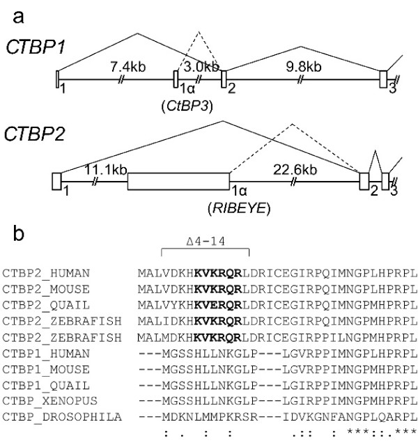
Background: CtBP1 and CtBP2 are transcriptional co-repressors that modulate the activity of a large number of transcriptional repressors via the recruitment of chromatin modifiers. Many CtBP-regulated proteins are involved in pathways associated with tumorigenesis, including TGF-beta and Wnt signalling pathways and cell cycle regulators such as RB/p130 and HDM2, as well as adenovirus E1A. CtBP1 and CtBP2 are highly similar proteins, although evidence is emerging that their activity can be differentially regulated, particularly through the control of their subcellular localisation. CtBP2s from diverse species contain a unique N-terminus, absent in CtBP1 that plays a key role in controlling the nuclear-cytoplasmic distribution of the protein.

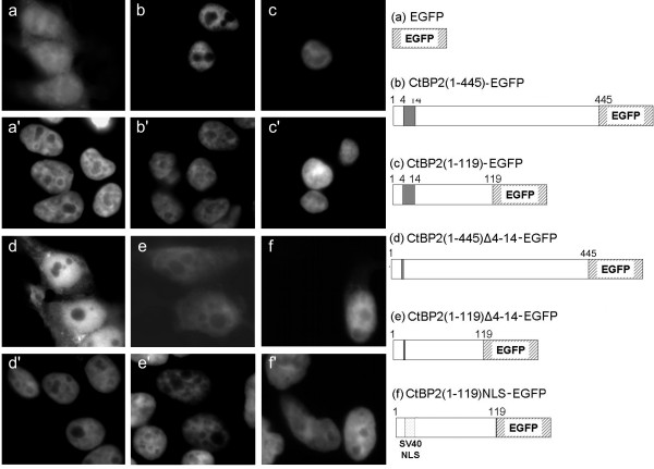
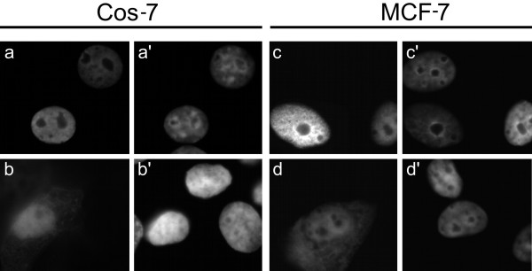
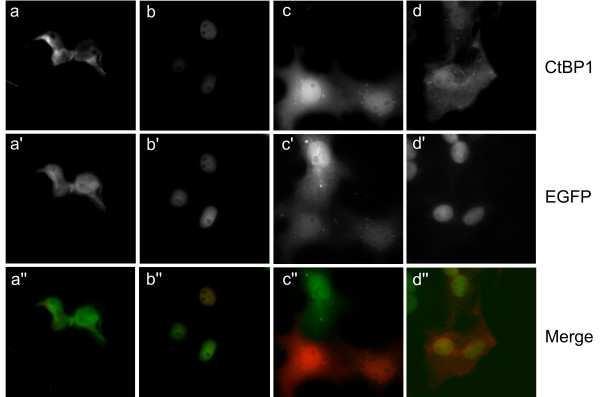
Results: Here we show that amino acids (a.a.) 4-14 of CtBP2 direct CtBP2 into an almost exclusively nuclear distribution in cell lines of diverse origins. Whilst this sequence contains similarity to known nuclear localisation motifs, it cannot drive nuclear localisation of a heterologous protein, but rather has been shown to function as a p300 acetyltransferase-dependent nuclear retention sequence. Here we define the region of CtBP2 required to co-operate with a.a. 4-14 to promote CtBP2 nuclear accumulation as being within a.a. 1-119. In addition, we show that a.a. 120-445 of CtBP2 can also promote CtBP2 nuclear accumulation, independently of a.a. 4-14. Finally, CtBP1 and CtBP2 can form heterodimers, and we show that the interaction with CtBP2 is one mechanism whereby CtBP1 can be recruited to the nucleus.

Conclusion: Together, these findings represent key distinctions in the regulation of the functions of CtBP family members that may have important implications as to their roles in development, and cell differentiation and survival.


Figures

Figure 1
Figure 1
CtBP gene structure and sequence comparison. (a) Genomic structure of the 5' end of human CTBP1 and CTBP2 genes. Published cDNA sequences were compared to human genome sequences using BLAST analysis. Solid lines show splicing of the major CTBP1 and CTBP2 transcripts, and dotted lines indicate the alternate splicing to generate CtBP1-S and RIBEYE. (b) ClustalW alignment of CtBP sequences from multiple higher organisms. Putative nuclear localisation signals are highlighted in bold type. The residues deleted in the Δ4–14 constructs are marked. Zebrafish have two ctbp2/ribeye loci [36].
Figure 2
Figure 2
Subcellular localisation of EGFP-tagged CtBP2 proteins in HEK 293 cells. Images are as follows: empty pEGFP-N1 vector (a); CtBP2(1–445)-EGFP (b); CtBP2(1–119)-EGFP (c); CtBP2(1–445)Δ4–14-EGFP (d); CtBP2(1–119)Δ4–14-EGFP (e); CtBP2(1–119)NLS-EGFP (f). Corresponding DAPI nuclear counterstain (a'-f'). A schematic diagram of the CtBP2-EGFP constructs is shown.
Figure 3
Figure 3
Subcellular localisation of CtBP proteins in HEK 293 cells, using myc-his-tagged constructs. Images are as follows: CtBP1(1–440)mh (a); CtBP2(1–445)mh (b); CtBP2(1–445)Δ4–14mh (c). Corresponding DAPI stains are shown in a'-c'.
Figure 4
Figure 4
Subcellular localisation of CtBP proteins in additional cell types. Expression patterns of CtBP1(1–440)mh, CtBP2(1–445)mh and CtBP2(1–445)Δ4–14mh in HeLa cells, (a,b,c respectively), Cos-7 cells (d,e,f respectively) and MCF-7 cells (g,h,i respectively). Corresponding DAPI images are shown in a'-i'.
Figure 5
Figure 5
Influence of PxDLS-containing proteins on CtBP2 subcellular localisation. Cos-7 and MCF-7 cells were transfected with either CtBP2(1–445)V72Rmh (a and c, respectively) or CtBP2(1–445)Δ4–14V72Rmh (b and d, respectively). Corresponding DAPI stains are shown in a'-d'.
Figure 6
Figure 6
Influence of CtBP2 on CtBP1 subcellular localisation. Cos-7 cells were co-transfected with 0.2 μg CtBP1(1–440)mh plus 0.2 μg of either control pEGFP-N1 (a,a',a"), CtBP2(1–445)-EGFP (b,b',b"), CtBP2(1–445)Δ4–14-EGFP (c,c',c"); or CtBP2(1–119)-EGFP (d,d',d"). a,b,c,d show localisation of CtBP1(1–440)mh. a',b',c',d' show localisation of EGFP fusion proteins. a",b",c",d" are merges of the CtBP1 and EGFP images.

References

    1. Boyd JM, Subramanian T, Schaeper U, La Regina M, Bayley S, Chinnadurai G. A region in the C-terminus of adenovirus 2/5 E1a protein is required for association with a cellular phosphoprotein and important for the negative modulation of T24-ras mediated transformation, tumorigenesis and metastasis. Embo J. 1993;12:469–478. - PMC - PubMed
    1. Shi Y, Sawada J, Sui G, Affar el B, Whetstine JR, Lan F, Ogawa H, Luke MP, Nakatani Y. Coordinated histone modifications mediated by a CtBP co-repressor complex. Nature. 2003;422:735–738. doi: 10.1038/nature01550. - DOI - PubMed
    1. Koipally J, Georgopoulos K. Ikaros interactions with CtBP reveal a repression mechanism that is independent of histone deacetylase activity. J Biol Chem. 2000;275:19594–19602. doi: 10.1074/jbc.M000254200. - DOI - PubMed
    1. Sewalt RG, Gunster MJ, van der Vlag J, Satijn DP, Otte AP. C-Terminal binding protein is a transcriptional repressor that interacts with a specific class of vertebrate Polycomb proteins. Mol Cell Biol. 1999;19:777–787. - PMC - PubMed
    1. Kim JH, Cho EJ, Kim ST, Youn HD. CtBP represses p300-mediated transcriptional activation by direct association with its bromodomain. Nat Struct Mol Biol. 2005;15:432–438. - PubMed

Publication types

MeSH terms