

APOBEC-1 and AID are nucleo-cytoplasmic trafficking proteins but APOBEC3G cannot traffic
- PMID: 16999936
- PMCID: PMC1847397
- DOI: 10.1016/j.bbrc.2006.09.032
APOBEC-1 and AID are nucleo-cytoplasmic trafficking proteins but APOBEC3G cannot traffic
Abstract
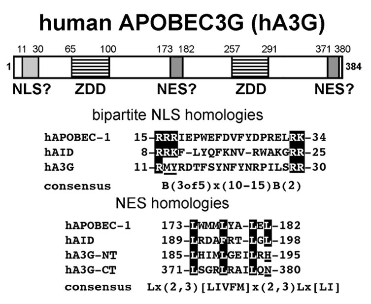
Human APOBEC3G (hA3G) is a member of the APOBEC-1 related protein (ARP) family of cytidine deaminases. hA3G functions as a natural defense against endogenous retrotransposons and a multitude of retroviruses, most notably human immunodeficiency virus type 1 (HIV-1). Nothing is known about the cellular function of hA3G, however, upon HIV-1 infection hA3G functions as an antiviral factor by mutating viral single-stranded DNA during reverse transcription. Whereas homologous deaminases such as APOBEC-1 and AID act on RNA and DNA, respectively, in the cell nucleus, hA3G mutagenic activity appears to be restricted to the cytoplasm. We demonstrate that hA3G is not a nucleo-cytoplasmic shuttling protein like APOBEC-1 and AID, but is strongly retained in the cytoplasm through a mechanism that involves both the N and C-terminal regions of the protein.
Figures


Similar articles
-
Nuclear Exclusion of the HIV-1 host defense factor APOBEC3G requires a novel cytoplasmic retention signal and is not dependent on RNA binding.J Biol Chem. 2008 Mar 21;283(12):7320-7. doi: 10.1074/jbc.M708567200. Epub 2007 Dec 28. J Biol Chem. 2008. PMID: 18165230
-
Functional analysis of the two cytidine deaminase domains in APOBEC3G.Virology. 2011 Jun 5;414(2):130-6. doi: 10.1016/j.virol.2011.03.014. Epub 2011 Apr 13. Virology. 2011. PMID: 21489586
-
APOBEC deaminases as cellular antiviral factors: a novel natural host defense mechanism.Med Sci Monit. 2006 May;12(5):RA92-8. Med Sci Monit. 2006. PMID: 16641889 Review.
-
Cytidine deamination and resistance to retroviral infection: towards a structural understanding of the APOBEC proteins.Virology. 2005 Apr 10;334(2):147-53. doi: 10.1016/j.virol.2005.01.038. Virology. 2005. PMID: 15780864 Review.
-
Activation-induced cytidine deaminase shuttles between nucleus and cytoplasm like apolipoprotein B mRNA editing catalytic polypeptide 1.Proc Natl Acad Sci U S A. 2004 Feb 17;101(7):1975-80. doi: 10.1073/pnas.0307335101. Epub 2004 Feb 9. Proc Natl Acad Sci U S A. 2004. PMID: 14769937 Free PMC article.
Cited by
-
Functions and consequences of AID/APOBEC-mediated DNA and RNA deamination.Nat Rev Genet. 2022 Aug;23(8):505-518. doi: 10.1038/s41576-022-00459-8. Epub 2022 Mar 7. Nat Rev Genet. 2022. PMID: 35256818 Free PMC article. Review.
-
RNA binding to APOBEC deaminases; Not simply a substrate for C to U editing.RNA Biol. 2017 Sep 2;14(9):1153-1165. doi: 10.1080/15476286.2016.1259783. Epub 2016 Nov 21. RNA Biol. 2017. PMID: 27869537 Free PMC article. Review.
-
The mechanisms regulating the subcellular localization of AID.Nucleus. 2010 Jul-Aug;1(4):325-31. doi: 10.4161/nucl.1.4.12107. Epub 2010 Mar 31. Nucleus. 2010. PMID: 21327080 Free PMC article.
-
Commentary on "Poor evidence for host-dependent regular RNA editing in the transcriptome of SARS-CoV-2".J Appl Genet. 2022 May;63(2):423-428. doi: 10.1007/s13353-022-00688-x. Epub 2022 Mar 12. J Appl Genet. 2022. PMID: 35279801 Free PMC article.
-
The dimerization domain of HIV-1 viral infectivity factor Vif is required to block virion incorporation of APOBEC3G.Retrovirology. 2007 Nov 24;4:81. doi: 10.1186/1742-4690-4-81. Retrovirology. 2007. PMID: 18036235 Free PMC article.
References
-
- Wedekind JE, Dance GS, Sowden MP, Smith HC. Messenger RNA editing in mammals: new members of the APOBEC family seeking roles in the family business. Trends Genet. 2003;19:207–216. - PubMed
-
- Sheehy AM, Gaddis NC, Choi JD, Malim MH. Isolation of a human gene that inhibits HIV-1 infection and is suppressed by the viral Vif protein. Nature. 2002;418:646–650. - PubMed
-
- Yu Q, Konig R, Pillai S, Chiles K, Kearney M, Palmer S, Richman D, Coffin JM, Landau NR. Single-strand specificity of APOBEC3G accounts for minus-strand deamination of the HIV genome. Nat Struct Mol Biol. 2004;11:435–442. - PubMed
-
- Yu X, Yu Y, Liu B, Luo K, Kong W, Mao P, Yu XF. Induction of APOBEC3G ubiquitination and degradation by an HIV-1 Vif-Cul5-SCF complex. Science. 2003;302:1056–1060. - PubMed
Publication types
MeSH terms
Substances
Grants and funding
LinkOut - more resources
Full Text Sources
Other Literature Sources
Miscellaneous
