Comparative Study
. 2006 Dec;26(23):8901-13.
doi: 10.1128/MCB.01156-06. Epub 2006 Sep 25.

Regulation of p53 localization and activity by Ubc13

Affiliations
Comparative Study

Regulation of p53 localization and activity by Ubc13

Aaron Laine et al. Mol Cell Biol. 2006 Dec.

Abstract

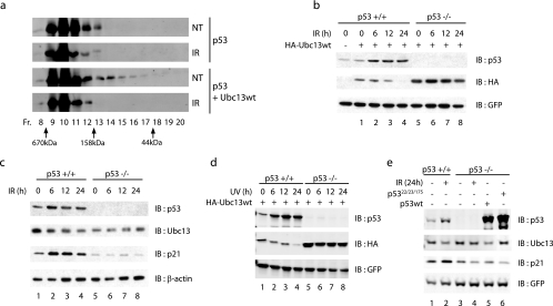
The abundance and activity of p53 are regulated largely by ubiquitin ligases. Here we demonstrate a previously undisclosed regulation of p53 localization and activity by Ubc13, an E2 ubiquitin-conjugating enzyme. While increasing p53 stability, Ubc13 decreases p53 transcriptional activity and increases its localization to the cytoplasm, changes that require its ubiquitin-conjugating activity. Ubc13 elicits K63-dependent ubiquitination of p53, which attenuates Hdm2-induced polyubiquitination of p53. Ubc13 association with p53 requires an intact C-terminal domain of p53 and is markedly stronger with a p53 mutant that cannot tetramerize. Expression of Ubc13 in vivo increases the pool of monomeric p53, indicating that Ubc13 affects tetramerization of p53. Significantly, wild-type but not mutant Ubc13 is associated with polysomes and enriches p53 within this fraction. In response to DNA damage, Ubc13 is no longer capable of facilitating p53 monomerization, in part due to a decrease in its own levels which is p53 dependent. Our findings point to a newly discerned mechanism important in the regulation of p53 organization, localization, and activity by Ubc13.


Figures

FIG. 1.
FIG. 1.
Ubc13 increases p53 levels. (a) Ubc13 stabilizes exogenous p53. HA-Ubc13 (2 μg) was cotransfected with 0.2 μg p53 into H1299 cells, and Western blot analysis was performed on whole-cell extracts (40 μg) with anti-p53 monoclonal antibody (DO-1) and anti-HA antibody. (b) Ubc13 requires its conjugating activity to stabilize endogenous p53. U2OS cells were transfected with increasing amounts of either wild-type (HA-Ubc13wt) or mutant (HA-Ubc13ca) Ubc13. p53 levels were analyzed by immunoblotting (IB). (c) Ubc13 expression does not increase p53 mRNA levels. mRNA was prepared from HCT116 p53+/+ cells expressing either the Ubc13 wild type or the C83A mutant. cDNA was generated and then used to perform reverse transcription PCR (20 cycles). The levels of transcript for p53, Ubc13, and GAPDH (glyceraldehyde-3-phosphate dehydrogenase) were assessed. (d) Ubc13 expression does not lead to the stabilization of the short-lived protein c-Myc. HCT116 p53+/+ cells were transfected with 2 μg of the HA-Ubc13 wild type or the C83A mutant. Cells were harvested, and the levels of c-Myc were determined by immunoblotting.
FIG. 2.
FIG. 2.
Ubc13 increases p53 stability by attenuating Mdm2-mediated degradation. (a) Ubc13 increases p53 half-life. HCT116 p53+/+ cells were transfected with either 2 μg empty plasmid or HA-Ubc13. Cells were treated with cycloheximide (CHX) (30 μg/ml) and harvested at the indicated times. p53 levels were analyzed by Western blotting with the DO-1 antibody. p53 levels were quantified and normalized relative to GFP levels. (b) Ubc13 attenuates Hdm2's ability to degrade p53. Whole-cell lysates from p53−//mdm2−/ MEFs that had been transfected with 0.2 μg p53 together with 2 μg Hdm2 and/or 1 μg wild-type (HA-Ubc13wt) or mutant (HA-Ubc13ca) Ubc13 were analyzed by Western blotting for p53 (top), HA (middle), or GFP (bottom). (c) Ubc13 does not compete with Hdm2 for binding to p53. H1299 cells were transfected with 1 μg p53, 2 μg Hdm2, and/or increasing amounts of WT Flag-Ubc13. Cells were treated with MG132 to avoid degradation of p53 upon expression of Hdm2 (40 μM for 4 h) before cells were harvested and p53 was immunoprecipitated using anti-p53 polyclonal antibodies (FL-393). Immunoprecipitates (IP) were separated by SDS-PAGE, and the amount of associated Hdm2 was assessed by Western blotting. Whole-cell extracts (WCE) were analyzed for relative levels of the indicated constructs. (d) Ubc13 attenuates Hdm2's ability to ubiquitinate p53 in vivo. p53−//mdm2−/ MEFs were cotransfected with 1.5 μg p53, 3 μg His-ubiquitin, 3 μg Hdm2, and 2 μg of the Ubc13 WT or the C83A mutant. Cells were treated with MG132 (40 μM) for 6 h before being harvested under denaturing conditions using 6 M guanidinium hydrochloride, and ubiquitinated proteins were pulled down by the addition of Ni2+-nitrilotriacetic acid agarose. The level of p53 ubiquitination was assessed by Western blotting with a mixture of anti-p53 monoclonal antibodies (DO-1, pAb421, and pAb241). The p53 forms conjugated with ubiquitin are indicated. *, monoubiquitinated form of p53; ←, diubiquinated form of p53. A fraction of the cell pellet was lysed in parallel using radioimmunoprecipitation assay buffer and used to determine the levels of p53, Hdm2, and Ubc13. IB, immunoblotting.
FIG. 3.
FIG. 3.
Ubc13 interacts with p53. (a) p53 interacts with Ubc13 in vitro. A complex between GST-Ubc13 and either His-MMS or His-Uev1A was preformed by incubating the corresponding proteins for 16 h at 4°C. After a wash, in vitro-transcribed and -translated p53 that had been radiolabeled with [35S]methionine was incubated with the preformed E2 complex for 4 h at 4°C. Samples were then separated by SDS-PAGE, stained with Coomassie blue, and exposed to film. (b) p53 directly interacts with Ubc13/Uev1A in vitro. Bacterially expressed and purified His-p53 and His-Ubc13/Uev1A (Boston Biochem) were incubated in 1% BSA binding buffer. The associated complex was then immunoprecipitated (IP) by using anti-p53 antibodies (DO-1) and protein G beads, washed, and then separated by SDS-PAGE. Normal mouse immunoglobulin G (NMIgG) was used as a control for the immunoprecipitation. (c) p53 interacts with Ubc13 in vivo. Whole-cell extracts (WCE) (2 mg) from HCT116 p53+/+ or p53−/− cells were immunoprecipitated using anti-Ubc13 monoclonal antibody. Levels of Ubc13-associated p53 were assessed by Western blotting using the DO-1 antibody. (d) Ubc13 interacts with p53 in vivo. HCT116 p53+/+ cells were harvested, and p53 was immunoprecipitated from 2 mg of whole-cell lysates by use of the DO-1 antibody. The level of associated Ubc13 was determined by Western blotting using anti-Ubc13 monoclonal antibodies (Zymed). Normal mouse immunoglobulin G was used as a control for immunoprecipitation. IB, immunoblotting.
FIG. 4.
FIG. 4.
Ubc13 regulates p53 ubiquitination. (a and b) Ubc13 regulates p53 ubiquitination in vivo. p53−//mdm2−/ MEFs were transfected and/or infected with the indicated constructs. Cells were harvested under denaturing conditions, and then proteins modified by His-tagged ubiquitin were pulled down by using Ni2+ agarose beads. The levels of ubiquitinated p53 were determined by Western blotting with a mixture of anti-p53 monoclonal antibodies (DO-1, pAb421, and pAb241). The p53 forms conjugated with ubiquitin are indicated. *, monoubiquitinated form of p53; ←, diubiquinated form of p53. Densitometric analysis was performed on the level of p53 ubiquitination with the values shown in panel b. (c) Mutant ubiquitin decreases levels of monoubiquitinated p53. His-tagged wild-type ubiquitin (Ubwt) or ubiquitin where either lysine 48 (UbK48R) or lysine 63 (UbK63R) was mutated to arginine (3 μg) was used for an in vivo ubiquitination reaction in p53−//mdm2−/ MEFs. (d) Ubc13 requires lysine 63 of ubiquitin to stabilize p53. p53−//mdm2−/ MEFs were transfected with the indicated constructs. Twenty-four hours after transfection, cells were harvested and protein levels determined by immunoblotting (IB).
FIG. 5.
FIG. 5.
Ubc13 highly associates with monomeric p53. The C-terminal region of p53 is required for Ubc13 interaction. H1299 cells were cotransfected with 2 μg wild-type (HA-Ubc13wt) or mutant (HA-Ubc13ca) Ubc13 and WT p53, p53 whose oligomerization domain had been deleted (the Δ325-354 mutant), p53 whose C-terminal domain had been deleted (the Δ363-393 mutant), or p53 in which several C-terminal lysines were mutated to arginine (the 7KR mutant). Whole-cell extracts (WCE) (1 mg) were used to immunoprecipitate (IP) Ubc13 with anti-HA antibody. Levels of associated p53 were determined by Western blotting with anti-p53 polyclonal antibody. IB, immunoblotting.
FIG. 6.
FIG. 6.
Ubc13 affects p53 oligomerization status. (a) Expression of Ubc13 increases monomeric levels of p53. H1299 cells were transfected with 1.5 μg p53 or p53 and 2 μg WT Ubc13, and then equal amounts of cellular lysate (400 μg) were fractionated by fast-performance liquid chromatography (FPLC) using a Sephadex 200 (30/300 GL) column. The fractions (Fr.) were then analyzed to determine p53 levels by Western blotting with the DO-1 antibody. The molecular masses of the fractions were determined by running a molecular mass standard in parallel. (b) Mutant Ubc13 increases the oligomerization of p53. H1299 cells were transfected with p53, p53 and WT Ubc13, or p53 and the Ubc13 C83A mutant (Ubc13ca). Cellular lysates (400 μg) were fractionated by FPLC using a Sephadex 200 (30/300 GL) column. The fractions were then analyzed by Western blotting with DO-1 antibody to determine the position of p53. The molecular masses of the fractions were determined by running a molecular mass standard in parallel. (c) Ubc13 affects oligomerization of endogenous p53. WT or mutant Ubc13 was transfected in HCT116 p53+/+ cells, and then lysates were fractionated by FPLC.
FIG. 7.
FIG. 7.
Ubc13 affects p53 activity and localization. (a) Ubc13 promotes nuclear export of p53. p53−//mdm2−/ MEFs were transfected with the indicated constructs (100 ng p53 or 500 ng Ubc13). Some cells were treated with leptomycin B (LMB) (2 μM) for 4 h before fixation. Subsequently, cells were permeabilized and stained for p53 (using polyclonal antibody FL-393) and Ubc13 (using monoclonal anti-HA antibody). Nuclei were counterstained with DAPI (4′,6′-diamidino-2-phenylindole), and images were taken through a confocal microscope. One hundred cells were counted for each treatment in two separate experiments, and the numbers of cells with higher levels of p53 in the cytoplasm versus nucleus (gray bars) and higher levels of p53 in the nucleus versus cytoplasm (black bars) were scored. (b) Ubc13 expression decreases p53 transcriptional activity. p53−//mdm2−/ MEFs were transfected with pGL3-Bax luciferase plasmid along with 0.1 μg p53 and/or Ubc13 (0.2 μg or 0.4 μg). The level of transcriptional activation was monitored by determining luciferase activity and normalizing to an internal control, β-galactosidase activity. (c) Ubc13 decreases the transcriptional activation of p21 by p53. p53−//mdm2−/ MEFs were cotransfected with p21-luciferase, p53, Ubc13, and β-galactosidase (normalization control). Luciferase activity was monitored by using a luciferase assay system (Promega). Values were normalized to β-galactosidase activity. (d) Leptomycin B attenuates Ubc13-mediated inhibition of p53's transactivation activity. p53−//mdm2−/ MEFs were transfected with pGL3-Bax luciferase plasmid along with p53 and/or Ubc13 (0.4 μg). Indicated lanes were treated with leptomycin B (2 μM) for 4 h before harvesting. The level of transcriptional activation was monitored by determining luciferase activity and normalizing to an internal control, β-galactosidase activity.
FIG. 8.
FIG. 8.
Ubc13 affects p53 apoptotic activity. (a) Ubc13 attenuates p53-mediated apoptosis. Saos-2 cells were transfected with 1 μg p53 alone or with p53 and 2 μg wild-type (Ubc13wt) or mutant (Ubc13ca) Ubc13. Thirty-six hours after transfection, cells were harvested. Cell cycle profiles, determined by staining DNA with propidium iodide followed by fluorescence-activated cell sorter analysis, and percentages of the sub-G1 population are shown. (b) Ubc13 knockdown increases p53 activity. U2OS cells were infected with either pRetroSuper containing shRNA against Ubc13 or scrambled control. Seventy-two hours after infection, cells were harvested and protein levels were determined by Western blotting. (c) Ubc13 associates with polysomes and perturbs the distribution of ribosome bound p53 protein. Ribosomal pellets obtained from the vector-, Ubc13 C83A mutant-, and Ubc13 WT-transfected U2OS cells were loaded onto 10 to 40% continuous sucrose density gradients. The absorbance (Abs) profiles of the gradients at 254 nm are shown at the top of each panel. Distribution of Flag-Ubc13, p53, and ribosomal protein S6 (rpS6) was determined by immunoblotting (IB). Importantly, Flag-Ubc13 and p53 associate with heavier polysomes only in Flag-Ubc13wt-transfected cells (fractions 8, 9, and 10 and 7 and 8, respectively). Distribution of rRNA across the gradient is given at the bottom of each panel. 80S, monosome (monomeric ribosome).
FIG. 9.
FIG. 9.
p53 regulates Ubc13 stability. (a) IR attenuates Ubc13's activity on p53. H1299 cells were transfected with the indicated constructs. Cells were treated with γ-irradation (10 Gy) and then harvested 4 h later. Lysates were subjected to fractionation by gel filtration. (b) IR treatment decreases exogenous Ubc13 expression in a p53-dependent manner. HCT116 p53+/+ or p53−/− cells were transfected with WT Ubc13. Cells were then treated with γ-irradiation (10 Gy) and harvested at the indicated time points. (c) p53 is required for decreased level of endogenous Ubc13 after IR. HCT116 p53+/+ or p53−/− cells were treated with γ-irradiation (10 Gy) and then harvested at the indicated time points. (d) Ubc13 levels are decreased upon various stimuli. HCT116 p53+/+ or p53−/− cells were transfected with WT Ubc13. Cells were then treated with 30 J/m2 of UV irradiation and harvested at the indicated time points. (e) p53 transcriptional activity is required to decrease Ubc13 levels. HCT116 p53+/+ or p53−/− cells were transfected with WT or transcriptionally inactive p53. Cells were then treated with γ-irradiation (10 Gy) and harvested at the indicated time points, and endogenous Ubc13 levels were determined. NT, nontreated; Fr., fraction; IB, immunoblotting.

References

    1. Andersen, P. L., H. Zhou, L. Pastushok, T. Moraes, S. McKenna, B. Ziola, M. J. Ellison, V. M. Dixit, and W. Xiao. 2005. Distinct regulation of Ubc13 functions by the two ubiquitin-conjugating enzyme variants Mms2 and Uev1A. J. Cell Biol. 170:745-755. - PMC - PubMed
    1. Ashcroft, M., and K. H. Vousden. 1999. Regulation of p53 stability. Oncogene 18:7637-7643. - PubMed
    1. Brooks, C. L., and W. Gu. 2003. Ubiquitination, phosphorylation and acetylation: the molecular basis for p53 regulation. Curr. Opin. Cell Biol. 15:164-171. - PubMed
    1. Brummelkamp, T. R., R. Bernards, and R. Agami. 2002. Stable suppression of tumorigenicity by virus-mediated RNA interference. Cancer Cell 2:243-247. - PubMed
    1. Brusky, J., Y. Zhu, and W. Xiao. 2000. UBC13, a DNA-damage-inducible gene, is a member of the error-free postreplication repair pathway in Saccharomyces cerevisiae. Curr. Genet. 37:168-174. - PubMed

Publication types

Substances