. 2006 Dec;26(23):8914-27.
doi: 10.1128/MCB.02299-05. Epub 2006 Sep 25.

The microphthalmia-associated transcription factor Mitf interacts with beta-catenin to determine target gene expression

Affiliations

The microphthalmia-associated transcription factor Mitf interacts with beta-catenin to determine target gene expression

Alexander Schepsky et al. Mol Cell Biol. 2006 Dec.

Abstract

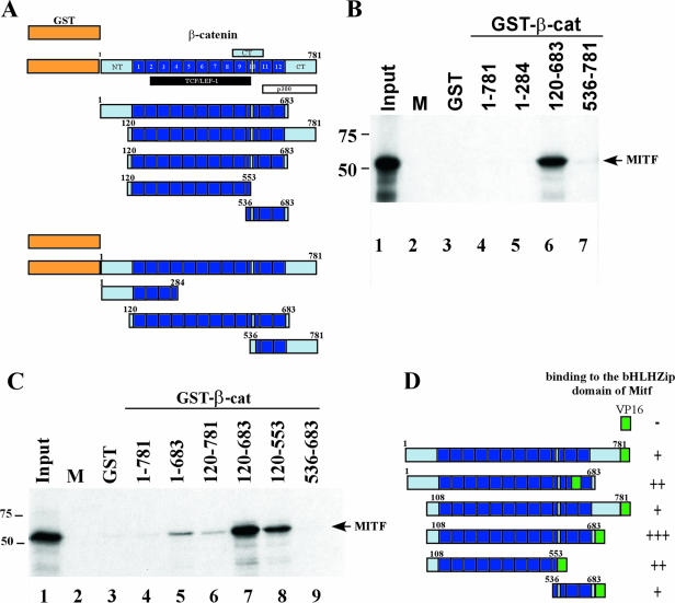
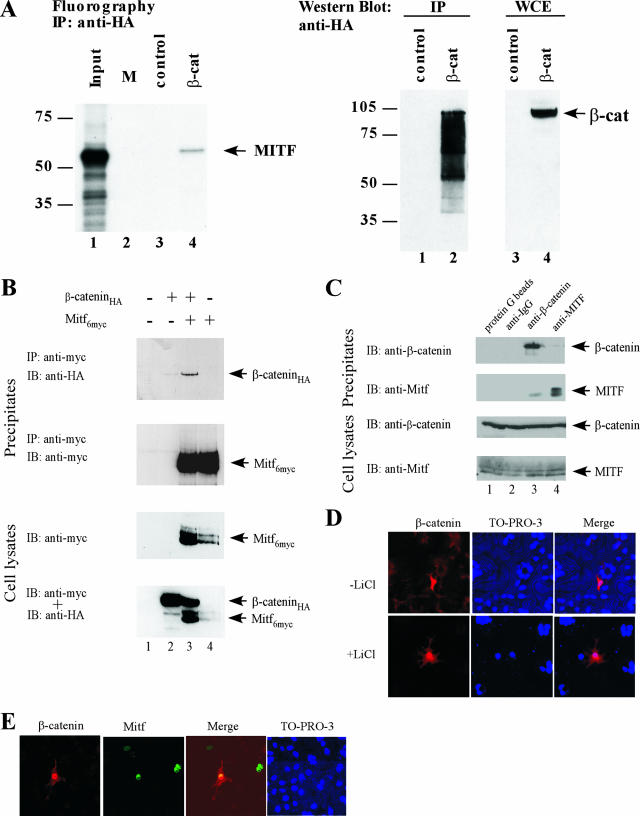
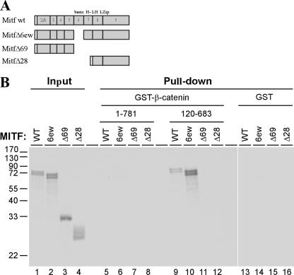
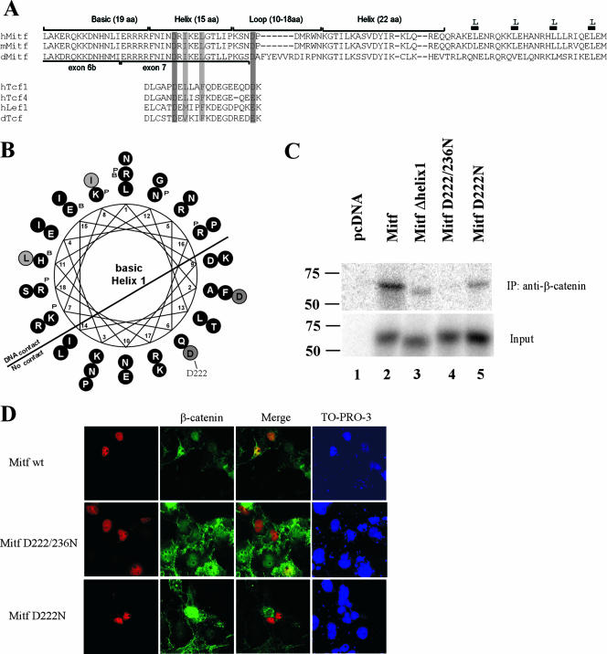
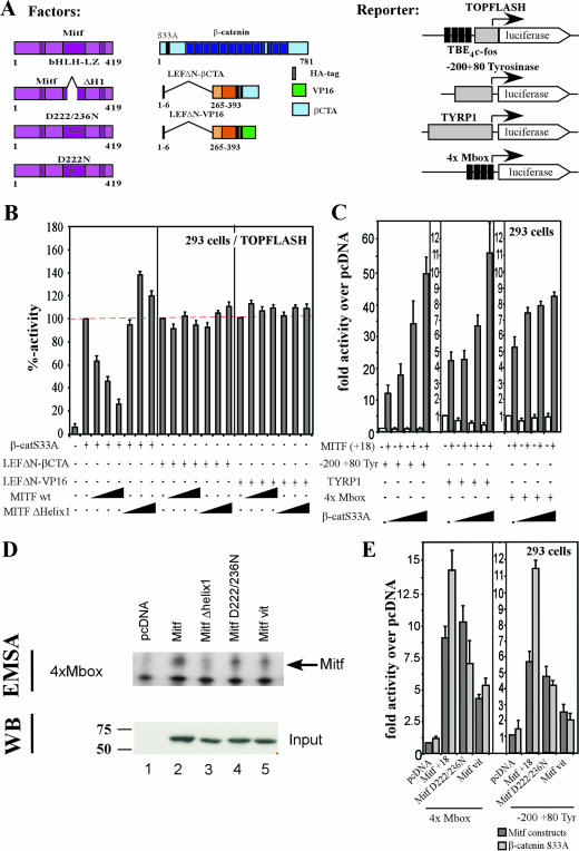
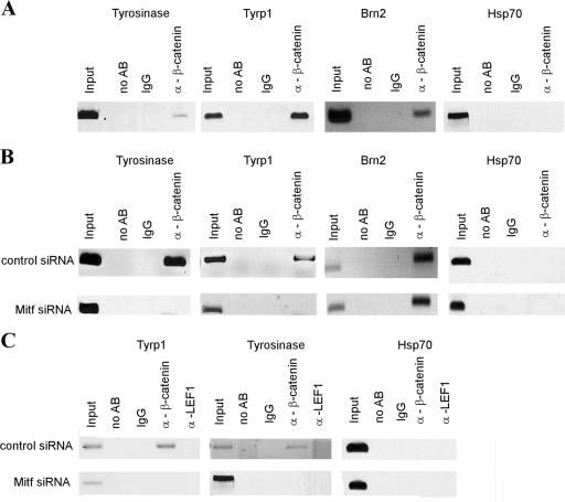
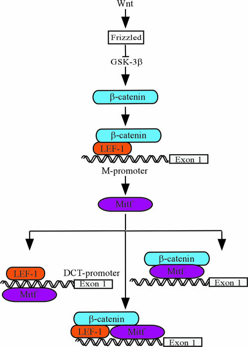
Commitment to the melanocyte lineage is characterized by the onset of expression of the microphthalmia-associated transcription factor (Mitf). This transcription factor plays a fundamental role in melanocyte development and maintenance and seems to be crucial for the survival of malignant melanocytes. Furthermore, Mitf has been shown to be involved in cell cycle regulation and to play important functions in self-renewal and maintenance of melanocyte stem cells. Although little is known about how Mitf regulates these various processes, one possibility is that Mitf interacts with other regulators. Here we show that Mitf can interact directly with beta-catenin, the key mediator of the canonical Wnt signaling pathway. The Wnt signaling pathway plays a critical role in melanocyte development and is intimately involved in triggering melanocyte stem cell proliferation. Significantly, constitutive activation of this pathway is a feature of a number of cancers including malignant melanoma. Here we show that Mitf can redirect beta-catenin transcriptional activity away from canonical Wnt signaling-regulated genes toward Mitf-specific target promoters to activate transcription. Thus, by a feedback mechanism, Mitf can diversify the output of canonical Wnt signaling to enhance the repertoire of genes regulated by beta-catenin. Our results reveal a novel mechanism by which Wnt signaling and beta-catenin activate gene expression, with significant implications for our understanding of both melanocyte development and melanoma.


Figures

FIG. 1.
FIG. 1.
Interactions between β-catenin and Mitf. (A) Schematic representation of the different β-catenin constructs used in the GST pull-down. Armadillo repeats are shown as numbered boxes. The binding domains of known β-catenin interacting partners, members of the Tcf/Lef1 transcription factor family, p300, and the C-terminal part of β-catenin are indicated below the full-length β-catenin protein. NT/CT: N-terminal and C-terminal tails of β-catenin, respectively. (B and C) Mapping the Mitf interaction domain in β-catenin. GST and GST-β-catenin fusion proteins were immobilized on glutathione-Sepharose beads, as indicated, and incubated with [35S]methionine-labeled Mitf. After extensive washing, material retained on the beads was analyzed by SDS-PAGE and fluorography, together with 10% of the input material. (D) Mapping the MITF interaction domain in β-catenin using the yeast two-hybrid assay. The different β-catenin constructs were tested for interactions with Mitf in a yeast two-hybrid assay. The different fragments used are outlined on the left, and their binding capacities are indicated on the right, as monitored by β-galactosidase activity: +++, high binding capacity; ++, good binding capacity; +, weak binding capacity; −, no binding capacity. β-cat, β-catenin; M, marker.
FIG. 2.
FIG. 2.
Coimmunoprecipitation of Mitf and β-catenin. Coimmunoprecipitation assays were performed to test the interaction between Mitf and β-catenin. (A) Cell lysates from HEK293 cells, transfected with an HA-tagged β-catenin construct, were incubated with in vitro translated and 35S-labeled Mitf, precipitated with an HA-specific antibody, and analyzed for the presence of Mitf using SDS-PAGE and radiography. β-catenin expression and immunoprecipitation were confirmed by Western blot analysis using a β-catenin-specific antibody. (B) HEK293 cells were cotransfected with HA-tagged β-catenin and c-Myc-tagged Mitf constructs. Forty-eight hours after transfection, cells were incubated with 40 mM LiCl for 4 h and harvested. Cell lysates were precipitated with a c-myc-specific antibody and analyzed for the presence of Mitf and β-catenin using SDS-PAGE and Western blot analysis. (C) Cell lysates of 501mel cells were precipitated with either β-catenin- or Mitf-specific antibodies and analyzed for the presence of Mitf and β-catenin using SDS-PAGE and Western blot analysis. (D) Effects of LiCl stimulation on β-catenin localization. COS7 cells were transfected with an expression vector for c-Myc-tagged β-catenin. Twenty-four hours posttransfection, the cells were stimulated with either 40 mM LiCl or NaCl for 3 h, fixed in 2% paraformaldehyde, permeabilized with PBS-Triton X-100, and labeled for β-catenin with a mouse anti-cMyc antibody (Abcam) and a secondary anti-mouse Alexa 568 antibody (Molecular Probes). (E) To analyze the nuclear localization patterns of Mitf and β-catenin, COS7 cells were cotransfected with a c-Myc-tagged β-catenin construct and a GFP-tagged Mitf construct. Twenty-four hours posttransfection, cells were fixed in 2% paraformaldehyde, permeabilized with PBS-Triton X-100, and labeled for β-catenin with an anti-cMyc antibody (Abcam) and a secondary anti-mouse Alexa 568 antibody (Molecular Probes). IP, immunoprecipitation; IB, immunoblotting; β-cat, β-catenin; WCE, whole-cell extract; IgG, immunoglobulin G; M, marker.
FIG. 3.
FIG. 3.
Mapping the β-catenin interaction domain in Mitf. (A) A schematic representation of the different Mitf deletion constructs used. Numbers indicate exons in the Mitf gene. basic HLH-LZip, region of the basic HLH leucine zipper domains. (B) Results of the GST pull-down assays. GST and GST-β-catenin fusion proteins were immobilized on glutathione-Sepharose beads as indicated and incubated with [35S]methionine-labeled Mitf and Mitf deletion mutants. After extensive washing, material retained on the beads was analyzed by SDS-PAGE and fluorography, together with 10% of the input material. WT, wild type.
FIG. 4.
FIG. 4.
Helix 1 of the Mitf bHLH domain provides critical contact points for the interaction with β-catenin. (A) Sequence comparison of the β-catenin binding domain of different Mitf homologues with known β-catenin binding domains of members of the Tcf/Lef1 transcription factor family. Protein alignments were carried out using ClustalW at the San Diego Supercomputer Center workbench (http://workbench.sdsc.edu/) and then further adjusted by eye. (B) To better visualize their potential functional importance, the positions of the conserved amino acids, helix 1, and the loop were also depicted in a helical wheel structure, using the sequence analysis program BIOEDIT. (C) To confirm the sequence analysis results, coimmunoprecipitation assays were performed. Cell lysates from HEK293 cells, transfected with an HA-tagged β-catenin construct, were incubated with the different in vitro translated and 35S-labeled Mitf deletion constructs, precipitated with an HA-specific antibody, and analyzed for the presence of the Mitf deletion construct using SDS-PAGE and radiography. β-Catenin expression and immunoprecipitation were confirmed by Western blot analysis using a β-catenin-specific antibody. (D) To analyze the importance of the D222 and D236 amino acids for the interaction with β-catenin, colocalization studies with the different Mitf mutant constructs were performed. COS7 cells were treated as before, and β-catenin was labeled with a goat anti-β-catenin antibody (Santa Cruz) and a secondary anti-goat Alexa 488 antibody (Molecular Probes). The nuclear localization of the different Mitf constructs was analyzed with a mouse anti-Mitf antibody and a secondary anti-mouse Alexa 568 antibody. IP, immunoprecipitation; wt, wild type.
FIG. 5.
FIG. 5.
Functional cooperation of Mitf and β-catenin. To test the functional relevance of the interactions, Mitf and β-catenin were tested in cotransfection assays in concert with β-catenin- or Mitf-specific reporter constructs. All the Mitf constructs contain the alternative 18-bp sequence just upstream of the basic domain (41a). (A) Schematic representation of the different constructs used. TBE4c-fos, four copies of the TCF binding element adjusted from the c-fos minimal promoter (gray box). (B) HEK293 cells were cotransfected with the TOPFLASH promoter construct and different activators, as indicated. The luciferase activity was measured, and the activity of the different activators was set as 100%. The influence of Mitf wild type (wt) and the Mitf Δhelix1 mutant construct on the activity of the different activators on the TOPFLASH promoter is shown as percent decrease. (C) HEK293 cells were cotransfected with either the −200/+80 tyrosinase promoter construct, the Tyrp-1 promoter construct, or a 4×M-box construct in concert with the different activators indicated. Mitf activity was measured as relative activity (n-fold) over pcDNA. (D) To determine whether the different Mitf mutant proteins are able to homodimerize and bind DNA, DNA band shift assays with in vitro translated proteins were performed. Binding specificity was confirmed by supershift with an Mitf-specific antibody. Input of equal amounts of the different proteins was confirmed by Western blotting. (E) To confirm the importance of the helix 1 region for the interaction with β-catenin, cotransfection studies with the different Mitf mutant constructs were performed. HEK293 cells were cotransfected with either the 4×M-box promoter construct or the −200/+80 tyrosinase construct and the different Mitf mutant constructs, with or without β-catenin.
FIG. 6.
FIG. 6.
Mitf recruits β-catenin to specific target genes. (A) Association of β-catenin with Mitf-regulated genes was analyzed by ChIP assays. 501mel melanoma cells were fixed in paraformaldehyde and harvested, and DNA was sheared by sonication. Cell lysates were precipitated with either a nonspecific antibody or a polyclonal β-catenin antibody. After precipitation, cross-links were reverted, and precipitated DNA was purified and analyzed via PCR for known Mitf binding sequences or for control sequences. (B) To confirm that the presence of β-catenin at the tyrosinase and Tyrp-1 promoters depends on Mitf-β-catenin interactions, Mitf was depleted using specific siRNAs prior to ChIP. (C) To confirm that the observed results were not dependent on a scaffold effect of Lef1, control ChIP assays with an anti-Lef1 antibody were performed on both control-depleted and Mitf-siRNA-depleted 501 cells. Cells were treated as above. IgG, immunoglobulin G; AB, antibody.
FIG. 7.
FIG. 7.
Model for the dynamic interactions between β-catenin and Mitf transcriptional activity. Mitf plays a key role at different stages of melanoblast and melanocyte development, probably by regulating different sets of target genes. To achieve this diversity, the activity of Mitf as a transcriptional activator is modulated either by regulating its protein level or by its engagement in changing and dynamic interactions with other factors in a highly regulated and promoter-specific manner. Moreover, signaling through the canonical Wnt signaling pathway provides several levels of regulation. Signaling through β-catenin activates Mitf expression. Depending on the protein levels of Mitf, Mitf can interact and cooperate either with Lef1 or β-catenin alone or in a complex with both to activate downstream targets.

References

    1. Aberle, H., S. Butz, J. Stappert, H. Weissig, R. Kemler, and H. Hoschuetzky. 1994. Assembly of the cadherin-catenin complex in vitro with recombinant proteins. J. Cell Sci. 107:3655-3663. - PubMed
    1. Bauer, A., O. Huber, and R. Kemler. 1998. Pontin52, an interaction partner of beta-catenin, binds to the TATA box binding protein. Proc. Natl. Acad. Sci. USA 95:14787-14792. - PMC - PubMed
    1. Behrens, J., B. A. Jerchow, M. Wurtele, J. Grimm, C. Asbrand, R. Wirtz, M. Kuhl, D. Wedlich, and W. Birchmeier. 1998. Functional interaction of an axin homolog, conductin, with beta-catenin, APC, and GSK3β. Science 280:596-599. - PubMed
    1. Bek, S., and R. Kemler. 2002. Protein kinase CKII regulates the interaction of beta-catenin with alpha-catenin and its protein stability. J. Cell Sci. 115:4743-4753. - PubMed
    1. Bienz, M. 2005. β-Catenin: a pivot between cell adhesion and Wnt signalling. Curr. Biol. 15:R64-R67. - PubMed

Publication types

MeSH terms

LinkOut - more resources