. 2006 Dec;26(24):9209-19.
doi: 10.1128/MCB.00478-06. Epub 2006 Sep 25.

Dimerization of the I kappa B kinase-binding domain of NEMO is required for tumor necrosis factor alpha-induced NF-kappa B activity

Affiliations

Dimerization of the I kappa B kinase-binding domain of NEMO is required for tumor necrosis factor alpha-induced NF-kappa B activity

Ralf B Marienfeld et al. Mol Cell Biol. 2006 Dec.

Abstract

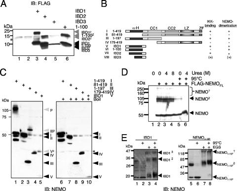
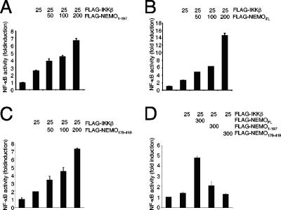
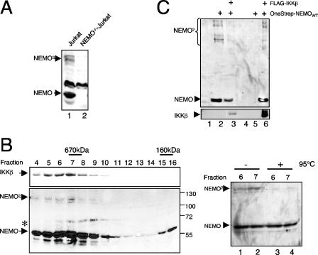
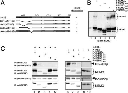
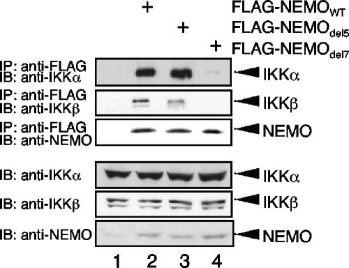
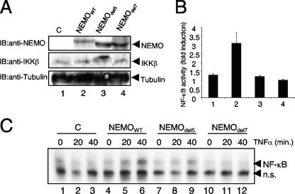
Previous studies have demonstrated that peptides corresponding to a six-amino-acid NEMO-binding domain from the C terminus of IkappaB kinase alpha (IKKalpha) and IKKbeta can disrupt the IKK complex and block NF-kappaB activation. We have now mapped and characterized the corresponding amino-terminal IKK-binding domain (IBD) of NEMO. Peptides corresponding to the IBD were efficiently recruited to the IKK complex but displayed only a weak inhibitory potential on cytokine-induced NF-kappaB activity. This is most likely due to the formation of sodium dodecyl sulfate- and urea-resistant NEMO dimers through a dimerization domain at the amino terminus of NEMO that overlaps with the region responsible for binding to IKKs. Mutational analysis revealed different alpha-helical subdomains within an amino-terminal coiled-coil region are important for NEMO dimerization and IKKbeta binding. Furthermore, NEMO dimerization is required for the tumor necrosis factor alpha-induced NF-kappaB activation, even when interaction with the IKKs is unaffected. Hence, our data provide novel insights into the role of the amino terminus of NEMO for the architecture of the IKK complex and its activation.


Figures

FIG. 1.
FIG. 1.
IKK-binding domains of NEMO are weak inhibitors of TNF-α-induced NF-κB activity. (A) A schematic representation of the NEMO peptides used in the analysis. (B) 293 cells were transiently transfected with expression vectors encoding either Xpress-tagged IKKα (lanes 1 to 5) or IKKβ (lanes 6 to 10) alone or with expression vectors for FLAG-tagged IBD1 (aa 1 to 120), IBD2 (aa 40 to 120) or IBD3 (aa 60 to 120). Forty-eight hours posttransfection, whole-cell extracts were prepared and the IBD:IKK complexes were precipitated using anti-FLAG beads. The resulting samples were subjected to an immunoblot (IB) analysis using anti-Xpress and anti-FLAG antibodies as indicated. A fraction of the extracts (10%) was subjected to immunoblot analysis with the indicated antibodies to check for the expression of the various ectopically expressed proteins. (C) A similar coimmunoprecipitation experiment was performed using F-IBD1 and NEMO1-100 in conjunction with Xpress-tagged IKKα or IKKβ. (D) 293 cells were transiently transfected with 3 μg of expression vectors encoding IBD1, IBD2, or NEMO1-100. Whole-cell extracts were prepared 48 h posttransfection and were used in immunoprecipitation experiments with anti-FLAG beads. The resulting immunoprecipitates were subjected to an immunoblot analysis using either the anti-IKKα/β antibody or the FLAG antibody (left part). As a control, a fraction of the whole-cell extracts was subjected to an anti-FLAG or an anti-IKKα/β immunoblot. The anti-IKKα/β antibody preferentially recognizes the IKKβ subunit. (E) 293 cells were transiently transfected with an NF-κB-dependent firefly luciferase reporter construct (200 ng) together with a renilla luciferase construct (30 ng) without or along with the indicated amounts of the different expression vectors for IBD1, IBD2, IBD3, and NEMO1-100. Twenty-four hours posttransfection, the cells were stimulated with TNF-α for four additional hours. Subsequently the cells were harvested, whole-cell extracts were prepared, and the firefly as well as the renilla luciferase activity was measured. The experiment was performed three times in parallel, and the mean value is shown. (F) 293 cells were transiently transfected with 0 or 3 μg F-IBD1 vector prior to immunoprecipitation experiments with anti-IKKα (upper part) or anti-IKKβ antibodies (lower part). The resulting immunoprecipitates (IP) were subjected to immunoblot analysis with the indicated antibodies. NRS, normal rabbit serum.
FIG. 2.
FIG. 2.
NEMO forms SDS-resistant dimers. (A) Whole-cell extracts from 293 cells transiently transfected with expression vectors for IBD1 (lane 3), IBD2 (lane 4), IBD3 (lane 5), or NEMO1-100 (lane 6) were subjected to an anti-NEMO immunoblot analysis. (B) Schematic representation of the different NEMO fragments used. (C) Anti-NEMO immunoblot of whole-cell extracts from exogenously expressed full-length NEMO (I in lanes 1 and 6), NEMO81-419 (II in lanes 2 and 7), NEMO1-197 (III in lanes 3 and 8), NEMO179-419 (IV in lanes 4 and 9), or IBD1 (V in lanes 5 and 10). The samples were either left untreated (left part) or boiled for 5 min (right part) prior to the separation by SDS-PAGE. The monomeric NEMO fragments are indicated by black, and the dimeric forms are indicated by gray arrows. *, unspecific bands. (D) Whole-cell extracts from 293 cells transiently transfected with an expression vector for FLAG-NEMO were either left untreated or were incubated with increasing concentrations of urea. Selected samples were heated (95°C) additionally. NEMO2, NEMO-containing high-molecular-weight protein complexes. (E) Ectopic expressed IBD1 or NEMO1-197 was either left untreated (lanes 1 to 2 and 5 to 6) or were subjected to a cross-linking procedure using 25 mM EGS (ethylene glycol bis[succinimidylsuccinate]) prior to an anti-NEMO immunoblot. An additional heating step was included as indicated. IB, immunoblot.
FIG. 3.
FIG. 3.
Amino-terminal NEMO dimerization is sufficient for the proximity-induced IKK activation. 293 cells were transiently transfected with a luciferase reporter construct controlled by a multimerized NF-κB-binding site in conjunction with a β-actin promoter-controlled renilla luciferase reporter construct, without or in combination with a small amount (25 ng) of an IKKβ-encoding expression vector. In addition, increasing amounts of an expression vector coding for (A) the amino terminus of NEMO (amino acids 1 to 197), (B) full-length NEMO, or (C) the carboxy terminus of NEMO (amino acids 179 to 419) were added. After 24 h, the cells were lysed in TNT and the activity of the firefly as well as the renilla luciferase was determined. A mean value of the corrected relative luciferase activity of two parallel experiments is shown. This experiment was performed three times with a similar result. (D) NEMO-deficient MEFs were transiently transfected with the indicated amounts of expression vectors for FLAG-tagged IKKβ, full-length NEMO, NEMO1-197, or NEMO179-419 in conjunction with the NF-κB-dependent firefly luciferase reporter construct and the β-actin promoter-dependent renilla luciferase reporter construct using the Lipofectamine 2000 reagent (Invitrogen).
FIG. 4.
FIG. 4.
Endogenous NEMO dimers are recruited to the IKK complex. (A) Dimer formation of endogenous NEMO. Fifty micrograms of whole-cell extract from wild-type Jurkat T cells (lane 1) or a NEMO-deficient Jurkat mutant (lane 2) was subjected to an anti-NEMO immunoblot analysis. (B) NEMO dimers comigrate with the IKK complex. S100 extracts from 4 × 10⁸ HeLa cells were fractionated on a Superose 6 column, and 6% of the indicated fractions was subjected to an anti-IKKβ (upper panel) or an anti-NEMO (lower panel) immunoblot analysis. The asterisk marks a nonspecific signal. As a control, 6% of the indicated fractions were either subjected to an additional heating step (right part, lanes 3 and 4) or was left untreated (lanes 1 and 2) prior to an anti-NEMO immunoblot analysis. (C) IKKβ interacts with NEMO monomers and dimers. 293 HEK cells were transiently transfected with 2 μg of expression vectors for FLAG-IKKβ and OneStrep-NEMO, as indicated, and the resulting whole-cell extracts were used for an anti-FLAG immunoprecipitation and subsequent anti-NEMO (upper part) or anti-IKKβ (lower part) immunoblot analysis.
FIG. 5.
FIG. 5.
NEMO dimer formation remains unaltered upon cell stimulation. Jurkat T cells were stimulated with TNF-α (A) or with PMA (B) for the indicated times, and whole-cell extracts were prepared. The resulting extracts were subjected to an anti-NEMO (upper panel) or an anti-IκBα immunoblot analysis (lower panel). As a control for the specificity of the NEMO signals, whole-cell extracts from NEMO-deficient Jurkat cells were included in the analysis (A and B, lanes 1). IB, immunoblot.
FIG. 6.
FIG. 6.
Different structural requirements for IKK binding and NEMO dimerization. (A) Schematic representation of the different deletion mutants of NEMO used. (B and C) 293 cells were either transiently transfected with expression vectors for the different FLAG-tagged NEMO variants alone (B) or in combination with expression vectors for Xpress-tagged IKKα or IKKβ (C). The resulting whole-cell extracts were subjected directly to an anti-NEMO immunoblot analysis (B) or to an anti-FLAG immunoprecipitation prior to anti-IKKα (C, left part), anti-IKKβ (C, right part), or anti-NEMO (C, both parts) immunoblot analysis. IB, immunoblot; IP, immunoprecipitate.
FIG. 7.
FIG. 7.
Reduced NEMO dimer formation and IKKβ binding by deletion of specific subdomains of the α-helical NEMO coiled-coil domain. (A) Schematic representation of the different NEMO variants used. (B) The NEMO dimer formation was monitored by an anti-NEMO immunoblot after transient transfection of 293 HEK cells with the indicated FLAG-tagged NEMO variants. (C) For the NEMO-IKKα interaction study, 2 μg of an Xpress-IKKα expression vector was either transfected alone or in combination with 1 μg of the indicated FLAG-NEMO variants. After 48 h, whole-cell extracts were prepared and an anti-FLAG immunoprecipitation experiment was performed. The resulting protein complexes were subjected to a Western blot analysis using the indicated antibodies. To control the expression of the different proteins, 10% of the lysate was used directly for an immunoblot. (D) An experiment similar to that described for panel C was performed after cotransfection of Xpress-IKKβ instead of IKKα. IB, immunoblot; IP, immunoprecipitate.
FIG. 8.
FIG. 8.
Interaction of NEMOdel5 and NEMOdel7 with endogenous IKK subunits. Whole-cell extracts from NEMO-deficient Jurkat T cells transiently transfected with the indicated FLAG-NEMO constructs were subjected to an anti-FLAG immunoprecipitation and subsequently anti-IKKα, anti-IKKβ, and anti-NEMO immunoblot analysis (upper part). As a control for the equal expression of the different IKK subunits, a fraction of the whole-cell extracts was subjected to immunoblot analysis with the indicated antibodies (lower part). IB, immunoblot; IP, immunoprecipitate.
FIG. 9.
FIG. 9.
Function analysis of the amino-terminal NEMO dimerization in vivo. (A) Anti-NEMO (upper panel), anti-IKKβ (middle panel), or anti-tubulin (lower panel) immunoblot analysis of 70 μg whole-cell extracts from the different retroviral transduced NEMO-deficient MEFs. (B) The different NEMO cell lines were transiently transfected with 400 ng of an NF-κB-dependent luciferase reporter construct and 30 ng of a β-actin-dependent renilla luciferase reporter. Eighteen hours posttransfection, the cells were either left untreated or stimulated with recombinant murine TNF-α (30 ng/ml) for five additional hours prior to the luciferase measurement. The ratio of the normalized values for stimulated versus unstimulated samples is depicted as fold induction. (C) NF-κB-specific electrophoretic mobility shift assay experiment with 10 μg nuclear proteins from the different untreated or TNF-α-stimulated NEMO cell lines. n.s., nonspecific signal. IB, immunoblot; C, control.

Similar articles

Cited by

References

    1. Agou, F., G. Courtois, J. Chiaravalli, F. Baleux, Y. M. Coic, F. Traincard, A. Israel, and M. Veron. 2004. Inhibition of NF-kappa B activation by peptides targeting NEMO oligomerization. J. Biol. Chem. 279:54248-54257. - PubMed
    1. Agou, F., F. Traincard, E. Vinolo, G. Courtois, S. Yamaoka, A. Israel, and M. Veron. 2004. The trimerization domain of NEMO is composed of the interacting C-terminal CC2 and LZ coiled-coil subdomains. J. Biol. Chem. 279:27861-27869. - PubMed
    1. Agou, F., F. Ye, S. Goffinont, G. Courtois, S. Yamaoka, A. Israel, and M. Veron. 2002. NEMO trimerizes through its coiled-coil C-terminal domain. J. Biol. Chem. 277:17464-17475. - PubMed
    1. Brummelkamp, T. R., S. M. Nijman, A. M. Dirac, and R. Bernards. 2003. Loss of the cylindromatosis tumour suppressor inhibits apoptosis by activating NF-kappaB. Nature 424:797-801. - PubMed
    1. Carter, R. S., K. N. Pennington, B. J. Ungurait, and D. W. Ballard. 2003. In vivo identification of inducible phosphoacceptors in the IKKgamma/NEMO subunit of human IkappaB kinase. J. Biol. Chem. 278:19642-19648. - PubMed

Publication types

MeSH terms

LinkOut - more resources