. 2006 Dec;26(23):8953-63.
doi: 10.1128/MCB.00506-06. Epub 2006 Sep 25.

The Ral/exocyst effector complex counters c-Jun N-terminal kinase-dependent apoptosis in Drosophila melanogaster

Affiliations

The Ral/exocyst effector complex counters c-Jun N-terminal kinase-dependent apoptosis in Drosophila melanogaster

Maria Balakireva et al. Mol Cell Biol. 2006 Dec.

Abstract

Ral GTPase activity is a crucial cell-autonomous factor supporting tumor initiation and progression. To decipher pathways impacted by Ral, we have generated null and hypomorph alleles of the Drosophila melanogaster Ral gene. Ral null animals were not viable. Reduced Ral expression in cells of the sensory organ lineage had no effect on cell division but led to postmitotic cell-specific apoptosis. Genetic epistasis and immunofluorescence in differentiating sensory organs suggested that Ral activity suppresses c-Jun N-terminal kinase (JNK) activation and induces p38 mitogen-activated protein (MAP) kinase activation. HPK1/GCK-like kinase (HGK), a MAP kinase kinase kinase kinase that can drive JNK activation, was found as an exocyst-associated protein in vivo. The exocyst is a Ral effector, and the epistasis between mutants of Ral and of msn, the fly ortholog of HGK, suggest the functional relevance of an exocyst/HGK interaction. Genetic analysis also showed that the exocyst is required for the execution of Ral function in apoptosis. We conclude that in Drosophila Ral counters apoptotic programs to support cell fate determination by acting as a negative regulator of JNK activity and a positive activator of p38 MAP kinase. We propose that the exocyst complex is Ral executioner in the JNK pathway and that a cascade from Ral to the exocyst to HGK would be a molecular basis of Ral action on JNK.


Figures
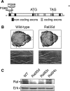
FIG. 1.
FIG. 1.
Characteristics of Ral mutants. (A) Schematic representation of the Ral locus and of the sites of insertion of transposons P{GawB} in strains PG69 and PG89, which were further excised to generate null and hypomorph mutants. (B) Scanning electron microscopy of complete nota (upper row) and fragments of nota (lower row) images documenting the loss-of-bristle phenotype of wild-type (left) and Ral35d (right) males. Higher magnification (lower row) shows that sockets are present and only shafts are missing, both for microchaetae and macrochaetae. (C) Western blot analysis of Ral protein expression in wild-type and in Ral35d, Ral83c, and Ral94c mutants. A total of 50 μg of protein from whole embryos was tested using antibodies against human RalB, which detects Drosophila Ral protein. Erk was used as a loading control.
FIG. 2.
FIG. 2.
Absence of shaft cells in sensory organs of Ral mutants. Wild-type and Ral35d nota were dissected at 30 h APF and 35 h APF, respectively. Both macrochaeta (A) and microchaeta (B) organs in wild-type flies were composed of four cells (visualized by Cut immunoreactivity [5]; green): one socket cell [large nuclear size and Su(H) immunoreactivity (21); red], a neuron (small nuclear size and HRP immunoreactivity [36]; blue), a shaft cell [large nuclear size and Su(H) negative], and a sheath cell (small nuclear size and HRP negative). In Ral mutants, the shaft cell was absent, and sensory organs were composed of three cells: the neuron, the socket, and the sheath cells. Each panel was obtained after merging about 10 horizontal confocal sections.
FIG. 3.
FIG. 3.
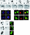
Shaft cell die by apoptosis in Ral mutants. (A and B) All four cells appear in sensory organs, and then one dies. Cell division in sensory organs of nota of live Ral35d; neuP72 UAS-H2B::YFP pupae was recorded by time-lapse microscopy (confocal for the macrochaeta lineage in panel A or standard epifluorescent for the microchaeta lineage in panel B). Time (hours:minutes) APF is indicated on each panel. Shaft cells (small arrows) were identified by size of their nuclei and their relative positions with respect to the other cells in the cluster. Shaft nuclear fragments (arrowheads) are observed at 26:36 h APF in the macrochaeta lineage (A) and at 33:53 h in the microchaeta lineage (B). Prior to fragmentation, shaft cells lost contact with the other cells in the cluster (26:28 h APF in macrochaeta in panel A; better views for both lineages are available in the films in the supplemental material). Cell movement was frequently associated with changes in the shape of the nucleus and DNA condensation that was observed as bright spots inside the nucleus. In panel A, each image corresponds to the merging of 10 horizontal confocal optical sections. The anterior is on the left. (C) Asymmetric distributions of Numb (Nb; green) and Baz (red) in wild-type and Ral35d dividing pI (upper row) and pIIa (lower row) cells. In both wild-type (WT; n = 10) and Ral35d (n = 17) mutant pI cells (identified by Senseless [Ss]; blue) at prometaphase, Numb was localized at the anterior cortex whereas Baz was localized at the posterior pI cell cortex. In both wild-type (n = 17) and Ral35d mutant (n = 21) pIIa cells, Numb was localized at the anterior cortex whereas Baz was enriched at the posterior pIIa cortex. The dividing pIIa cells were identified by cytoplasmic Cut staining and the presence of the pIIb daughter cells. In the lower row, the insets show the maximal projections of several confocal planes to illustrate the position of the dividing pIIa cells and the anterior pIIIb cells. Nota were dissected at 17 h APF to see pI division and at 21 to 22 h APF to see pIIa and pIIIb divisions. Scale bar, 5 μm. (D and E) The shaft cells die by apoptosis. Cell death was characterized by TUNEL staining performed on macrochaeta (upper row) and microchaeta (lower row) sensory organs at 25 h APF and 35 h APF, respectively. All four cells were detected by green fluorescence due to expression of H2B::YFP, and TUNEL-positive cells were detected by red fluorescence (Cy3-conjugated streptavidin). TUNEL-positive cells were identified as shaft cells by nuclear size and position. (E) Cell apoptosis was also visualized by the presence of an activated caspase-3 in one cell per organ in macrochaetae (upper row) and microchaetae (middle row), as opposed to wild-type organs (lower row). All four cells were detected by Cut immunoreactivity.
FIG. 4.
FIG. 4.
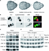
Ral brakes JNK, a conserved function in flies and human. (A) Ral behaves as a negative regulator of the JNK pathway in flies. Nomarsky microscopy of adult nota documents examples of genetic interactions between Ral mutants and mutants of the JNK pathway. The loss-of-bristle phenotype of Ral35d mutant flies (left image) was either suppressed (middle image) when the JNK pathways were down-regulated (as in the present example with a dominant negative allele of Jun expressed under the control the sca-Gal4 driver) or enhanced (as in the example of the right image, where the amount of MSN was increased under the control of the sca-Gal4 driver). Results of all tested interactions are presented in Table 1. (B) JNK is unduly activated in shaft cells of Ral mutants. lacZ expression under control of the Puckered promoter in the pucE69 mutant (49) is a reporter for JNK activity. lacZ-positive neurons are present in both wild-type and Ral mutant flies, but lacZ staining in shaft cells is observed only in Ral mutants. Cut staining reveals all four cells; HRP detects the neuron. (C and D) RalA inhibits basic JNK activation in human cells and impairs TNF-α activation of p38 MAP kinase. HeLa cells were transfected with control or RalA siRNAs. In panel C, at 72 h posttransfection, whole-cell lysates of attached cells (“attached”) or cells detached from plates with trypsin and maintained in suspension for 24 h (“suspension”) were prepared. Equivalent total protein was analyzed by sodium dodecyl sulfate-polyacrylamide gel electrophoresis for the indicated proteins (phospho-JNK corresponds to JNK1 and 2 phosphorylated on Thr183 and Tyr185; phospho-c-Jun corresponds to c-Jun phosphorylated on Ser73). JNK1/2 and ERK1/2 are shown as a loading control. In panel D, at 72 h posttransfection cells were treated with 10 ng/ml of TNF-α for the indicated times, and cell extracts were an analyzed for p38 MAP kinase phosphorylation as well as for RalA depletion. ERK1/2 is shown as a loading control.
FIG. 5.
FIG. 5.
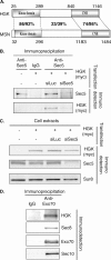
The exocyst complex interacts with HGK/MAP4K4, an activator of the JNK pathway. (A) Schematic representation of human and Drosophila MAP4K4 HGK and MSN. Domains are indicated as well as the percent identity/similarity between the different regions of the human and fly proteins. The global identity and similarity for the whole proteins are 43 and 53%, respectively. For the sake of simplicity, we show only isoform 1 of HGK (NP_004825). The difference between the three reported isoforms affects the regions flanking the kinase domain and the CNH domain. (B) Sec5 interacts with HGK. HeLa cells were transfected with the indicated plasmids and the indicated siRNAs. Whole-cell extracts were immunoprecipitated with the indicated antibodies. Myc-HGK was present in immunoprecipitates obtained with anti-Sec5 antibodies (58) in cells transfected with a plasmid expressing myc-HGK. This result was specific: when Sec5 protein was depleted by specific siRNA, less Sec5 and less myc-HGK were detected in the anti-Sec5 immunoprecipitate, showing that myc-HGK was not precipitated directly by the anti-Sec5 antibodies. (C) siRNAs against Sec5 are specific. The cells extracts used in panel B for immunoprecipitation were probed for the presence of the indicated proteins. The siRNA against Sec5 (siSec5) reduced strongly the amount of Sec5, while myc-HGK looks only marginally affected. (D) HGK is immunoprecipitated with the Exo70 subunit of the exocyst complex. NRK cells transfected with a plasmid expressing myc-HGK were lysed, and cell extracts were immunoprecipitated with the indicated antibodies. Immunoprecipitates were tested for the presence of other subunits of the exocyst and of HGK. HGK was detected in the immunoprecipitate with anti-Exo70. IgG, immunoglobulin G; siLuc, siRNA against luciferase.

Similar articles

Cited by

References

    1. Adachi-Yamada, T., M. Nakamura, K. Irie, Y. Tomoyasu, Y. Sano, E. Mori, S. Goto, N. Ueno, Y. Nishida, and K. Matsumoto. 1999. p38 mitogen-activated protein kinase can be involved in transforming growth factor β superfamily signal transduction in Drosophila wing morphogenesis. Mol. Cell. Biol. 19:2322-2329. - PMC - PubMed
    1. Agapova, L. S., J. L. Volodina, P. M. Chumakov, and B. P. Kopnin. 2004. Activation of Ras-Ral pathway attenuates p53-independent DNA damage G2 checkpoint. J. Biol. Chem. 279:36382-36389. - PubMed
    1. Bellaiche, Y., M. Gho, J. A. Kaltschmidt, A. H. Brand, and F. Schweisguth. 2001. Frizzled regulates localization of cell-fate determinants and mitotic spindle rotation during asymmetric cell division. Nat. Cell Biol. 3:50-57. - PubMed
    1. Bellaiche, Y., A. Radovic, D. F. Woods, C. D. Hough, M. L. Parmentier, C. J. O'Kane, P. J. Bryant, and F. Schweisguth. 2001. The partner of inscuteable/discs-large complex is required to establish planar polarity during asymmetric cell division in Drosophila. Cell 106:355-366. - PubMed
    1. Blochlinger, K., R. Bodmer, L. Y. Jan, and Y. N. Jan. 1990. Patterns of expression of cut, a protein required for external sensory organ development in wild-type and cut mutant Drosophila embryos. Genes Dev. 4:1322-1331. - PubMed

Publication types

MeSH terms

Substances