

Review
doi: 10.1084/jem.20061800.
Epub 2006 Sep 25.
Reinvigorating exhausted HIV-specific T cells via PD-1-PD-1 ligand blockade
Affiliations
- PMID: 17000870
- PMCID: PMC2118103
- DOI: 10.1084/jem.20061800
Item in Clipboard
Review
Reinvigorating exhausted HIV-specific T cells via PD-1-PD-1 ligand blockade
J Exp Med.
.
Abstract
The programmed death (PD)-1-PD-1 ligand (PD-L) pathway, which is part of the B7-CD28 family, consists of the PD-1 receptor and its two ligands PD-L1 and PD-L2. Engagement of PD-1 by its ligands inhibits immune responses, and recent work has shown that PD-1 is highly expressed on exhausted T cells during chronic lymphocytic choriomeningitis virus (LCMV) infection in mice. Blockade of this pathway reinvigorates the exhausted T cells, allowing them to expand and produce effector cytokines, raising the issue of whether this pathway has been exploited by a variety of viruses during chronic infection. New studies now extend these observations to HIV infection and human disease.
Figures
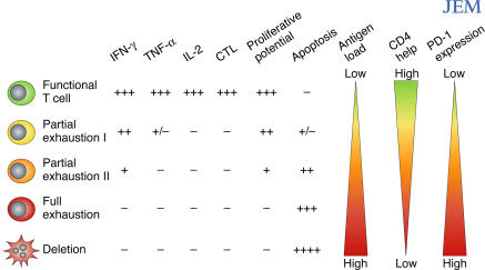
T cell exhaustion during chronic viral infections. Virus-specific CD8 T cells posses multiple functions including production of IFN-γ, TNF-α, IL-2, cytotoxicity, antigen-driven proliferation, and resistance to apoptosis. During chronic infections, functions can be exhausted. Exhaustion represents a spectrum from mild (Partial exhaustion I: little IL-2 and poor TNF-α and cytotoxicity) to moderate (Partial exhaustion II: modestly defective IFN-γ, cytotoxicity, and little IL-2 or TNF-α) to severe (Full exhaustion: lack of IFN-γ, TNF-α, IL-2, and cytotoxicity). Finally, physical deletion (apoptosis) of T cells occurs. Proliferative potential decreases concomitantly with the loss of other functions while apoptosis increases. Antigen and CD4 help strongly influence exhaustion; as antigen increases and/or CD4 help decreases, virus-specific T cells become more exhausted. Recent studies now identify the PD-1–PD-L pathway as a key regulator of exhaustion. Increased expression of PD-1 by virus-specific T cells, and PD-L1 by APCs, leads to more severe exhaustion during chronic viral infection.
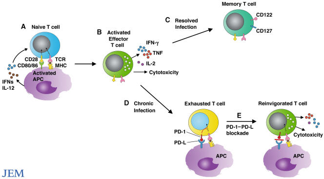
Reinvigorating exhausted T cells. (A) Microbial products and cytokines produced in response to microbes activate APCs and stimulate expression of CD80 and CD86. Engagement of CD28 by CD80/CD86 stimulates the expansion and differentiation of naive T cells into effector T cells. (B) Effector T cells eliminate the invading pathogens by secreting cytokines and killing infected cells. (C) Upon resolution of infection, effector T cells give rise to long-lived protective memory T cells. (D) However, during chronic infection, T cells lose function and the ability to proliferate and become functionally exhausted. Exhausted T cells express high levels of PD-1. (E) Blockade of interactions between PD-1 and its ligands can reinvigorate T cells to expand and regain effector functions, including cytokine production and cytolysis.
Comment on
-
PD-1 is a regulator of virus-specific CD8+ T cell survival in HIV infection.J Exp Med. 2006 Oct 2;203(10):2281-92. doi: 10.1084/jem.20061496. Epub 2006 Sep 5. J Exp Med. 2006. PMID: 16954372 Free PMC article.
References
-
- Okazaki, T., and T. Honjo. 2006. The PD-1-PD-L pathway in immunological tolerance. Trends Immunol. 27:195–201. - PubMed
-
- Greenwald, R.J., G.J. Freeman, and A.H. Sharpe. 2005. The B7 family revisited. Annu. Rev. Immunol. 23:515–548. - PubMed
-
- Latchman, Y., C.R. Wood, T. Chernova, D. Chaudhary, M. Borde, I. Chernova, Y. Iwai, A.J. Long, J.A. Brown, R. Nunes, et al. 2001. PD-L2 is a second ligand for PD-1 and inhibits T cell activation. Nat. Immunol. 2:261–268. - PubMed
-
- Brown, J.A., D.M. Dorfman, F.R. Ma, E.L. Sullivan, O. Munoz, C.R. Wood, E.A. Greenfield, and G.J. Freeman. 2003. Blockade of programmed death-1 ligands on dendritic cells enhances T cell activation and cytokine production. J. Immunol. 170:1257–1266. - PubMed
Publication types
MeSH terms
Substances
Grants and funding
LinkOut - more resources
Full Text Sources
Other Literature Sources
Medical
Research Materials
