. 2006 Sep 25;174(7):1107-17.
doi: 10.1083/jcb.200606056.

Flow-enhanced adhesion regulated by a selectin interdomain hinge

Affiliations

Flow-enhanced adhesion regulated by a selectin interdomain hinge

Jizhong Lou et al. J Cell Biol. .

Abstract

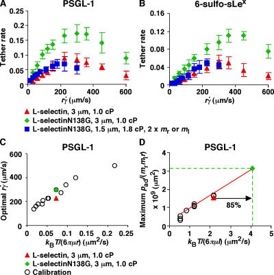
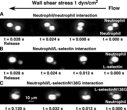
L-selectin requires a threshold shear to enable leukocytes to tether to and roll on vascular surfaces. Transport mechanisms govern flow-enhanced tethering, whereas force governs flow-enhanced rolling by prolonging the lifetimes of L-selectin-ligand complexes (catch bonds). Using selectin crystal structures, molecular dynamics simulations, site-directed mutagenesis, single-molecule force and kinetics experiments, Monte Carlo modeling, and flow chamber adhesion studies, we show that eliminating a hydrogen bond to increase the flexibility of an interdomain hinge in L-selectin reduced the shear threshold for adhesion via two mechanisms. One affects the on-rate by increasing tethering through greater rotational diffusion. The other affects the off-rate by strengthening rolling through augmented catch bonds with longer lifetimes at smaller forces. By forcing open the hinge angle, ligand may slide across its interface with L-selectin to promote rebinding, thereby providing a mechanism for catch bonds. Thus, allosteric changes remote from the ligand-binding interface regulate both bond formation and dissociation.


Figures

Figure 1.
Figure 1.
Selectin conformational changes regulated by an interdomain hinge. (A) X-ray structures of the lectin and EGF domains of P-selectin with a closed (left; PDB 1G1Q) and open angle (right; PDB 1G1S; Somers et al., 2000). The golden sphere at the top of the lectin domain represents a Ca2+ ion that is coordinated as part of the ligand-binding site. (B) X-ray (left) and MD-simulated (right) structures of the lectin and EGF domains of L-selectin with a closed (left) and open angle (right). The respective open- and closed-angle structures of P- and L-selectin align well. The boxed areas (left) highlight the putative hinge regions that are magnified in the insets. A hydrogen bond (dotted line) connects Tyr37 with Asn138, but not with Gly138. (C) RMSD between the corresponding backbone atoms from residues 121–156 (the EGF domain) of the simulated L-selectin structure and the crystal structures of closed-angle L-selectin (blue curve) or open-angle P-selectin (red curve) as a function of simulation time. The lectin domains were aligned by minimizing the RMSD between the backbone atoms from residues 1–120.
Figure 2.
Figure 2.
Effects of interdomain hinge flexibility on flow-enhanced tethering. (A and B) Tether rates of microspheres of indicated radii bearing either L-selectin or L-selectinN138G perfused in media of indicated viscosities were plotted against the product formula image, where r is the microsphere radius and formula image is the wall shear rate. The microspheres were coated with 750 molecules μm−2 of either L-selectin or L-selectinN138G, except in one case in B, where they were coated with 1,500 molecules μm−2 of L-selectinN138G (blue squares). The flow chamber floor in A was coated with PSGL-1 at either 120 (red triangles and green diamonds) or 240 (blue squares) molecules μm−2. The flow chamber floor in B was coated with a constant density of 6-sulfo-sLex. The data in A and B represent the mean ± the SD from three experiments. (C) Optimal formula image (peak locations of the p ad/(m r m l r) vs. formula image curves) versus microsphere diffusivity k B T/(6πμr) data for tethering to PSGL-1 (open circles; Yago et al., 2006) were replotted to provide calibration. The diffusivities of 3-μm radius microspheres bearing L-selectin (red triangle) or L-selectinN138G (green diamond) were plotted for comparison. (D) Maximum p ad/(m r m l r) versus molecular diffusivity k B T/(6πμl) data for tethering to PSGL-1 (open circles; unpublished data) were replotted to provide calibration, which assumed the characteristic length for molecular diffusion as l = 100 nm. The same l value was used for the L-selectin datum (red triangle), which matched the calibration curve well. The l value for L-selectinN138G is predicted to be smaller. Using the measured maximum p ad/(m r m l r) value, the L-selectinN138G datum point (green diamond) was located at the intercept of y = [p ad/(m r m l r)] max (green dashed horizontal line) and the extrapolation of the calibration curve (red line). The increased molecular diffusivity for L-selectinN138G could be calculated from the x-axis value of this datum point (green dashed vertical line).
Figure 3.
Figure 3.
Sliding–rebinding model for selectin–ligand interactions. The interdomain hinge is represented as a coiled spring. The yellow arrows indicate possible sequences of events. On the top left, a ligand binds to a selectin with a closed interdomain angle. A low applied force (f, short arrow) perpendicular to the binding interface favors ligand dissociation (bottom left). As applied force increases (f, long arrow), the equilibrium between the closed- and open-angle conformations shifts in favor of the open conformation (top right). This tilts the interface to align with the force direction, allowing the ligand to slide across the interface. Sliding allows new interactions to form or the original interactions to reform (rebinding). Eventually both old and new interactions break, and the ligand dissociates (bottom right).
Figure 4.
Figure 4.
Effects of interdomain hinge flexibility on catch bonds. (A and C) Interactions of L-selectin or L-selectinN138G with PSGL-1. (B and D) Interactions of L-selectin or L-selectinN138G with 6-sulfo-sLex. Bond lifetimes were measured by BFP (A and B) or by flow chamber (C and D) experiments (points), which were fitted by the sliding–rebinding model using Monte Carlo simulations (curves). The data in A and B represent the mean ± the SEM of ∼100 lifetime measurements. The data in C and D represent the mean ± the SD from five experiments. The fitting parameters areas follows: k +1 = 30 s−1; formula image; a = 0.2–0.4 Å, −f 1 = 5–40 pN; f 2 = 35–85 pN; and k +2 = 350–5,400 s−1.
Figure 5.
Figure 5.
Effects of interdomain hinge flexibility on flow-enhanced rolling. Microspheres bearing matched densities of L-selectin or L-selectinN138G were perfused through a flow chamber containing immobilized PSGL-1 (A, C, and E) or 6-sulfo-sLex (B, D, and F). Rolling parameters are presented as mean rolling velocities (A and B), mean stop time (C and D), or fractional stop time (E and F). The data in A and B represent the mean ± the SD from five experiments. The data in C–F represent analyses of thousands of rolling steps collected from 10–15 microspheres rolling for 1 s at each wall shear-stress for each pair of selectin–ligand interactions.
Figure 6.
Figure 6.
Effects of interdomain hinge flexibility on lifetimes of doublets of neutrophils or microspheres in a flow field. Neutrophils or mixtures of neutrophils with microspheres bearing L-selectin or L-selectinN138G were perfused through a flow chamber coated with HSA. Representative images of randomly colliding cells and/or microspheres were collected at 250 frames/s. A doublet formed by collision of two neutrophils (A) or of a neutrophil and an L-selectin microsphere (B), each dissociated within 0.028 s. In contrast, a doublet formed by the collision of a neutrophil and an L-selectinN138G microsphere (C) persisted for at least 0.1 s, until it flowed out of the field of view.
Figure 7.
Figure 7.
Quantification of neutrophil–microsphere aggregation in a flow field. Mixtures of neutrophils labeled with the red dye PKH26 and microspheres labeled with the green dye FITC were perfused through a flow chamber coated with HSA, as in Fig. 6. After exiting the flow chamber, the suspensions were fixed and analyzed by flow cytometry or by fluorescence microscopy. (A and D) Mixtures of neutrophils and L-selectin microspheres. (B and E) Mixtures of neutrophils and L-selectinN138G microspheres. (C and F) Mixtures of neutrophils and L-selectinN138G microspheres perfused in the presence of the anti–L-selectin mAb DREG-56. (A–C) Flow cytometry of ungated samples. The percentage of particles labeled with both dyes is listed in the top right quadrant. (D–F) Representative fluorescence micrographs. The data are representative of three independent experiments.

Comment in

References

    1. Ajzenberg, N., A.S. Ribba, G. Rastegar-Lari, D. Meyer, and D. Baruch. 2000. Effect of recombinant von Willebrand factor reproducing type 2B or type 2M mutations on shear-induced platelet aggregation. Blood. 95:3796–3803. - PubMed
    1. Alon, R., S.Q. Chen, K.D. Puri, E.B. Finger, and T.A. Springer. 1997. The kinetics of L-selectin tethers and the mechanics of selectin-mediated rolling. J. Cell Biol. 138:1169–1180. - PMC - PubMed
    1. Bell, G.I. 1978. Models for the specific adhesion of cells to cells: a theoretical framework for adhesion mediated by reversible bonds between cell surface molecules. Science. 200:618–627. - PubMed
    1. Doggett, T.A., G. Girdhar, A. Lawshe, J.L. Miller, I.J. Laurenzi, S.L. Diamond, and T.G. Diacovo. 2003. Alterations in the intrinsic properties of the GPIbα-VWF tether bond define the kinetics of the platelet-type von Willebrand disease mutation, Gly233Val. Blood. 102:152–160. - PubMed
    1. Dwir, O., G.S. Kansas, and R. Alon. 2000. An activated L-selectin mutant with conserved equilibrium binding properties but enhanced ligand recognition under shear flow. J. Biol. Chem. 275:18682–18691. - PubMed

Publication types