

The surface oxidation potential of human neuromelanin reveals a spherical architecture with a pheomelanin core and a eumelanin surface
- PMID: 17001010
- PMCID: PMC1595429
- DOI: 10.1073/pnas.0604010103
The surface oxidation potential of human neuromelanin reveals a spherical architecture with a pheomelanin core and a eumelanin surface
Abstract
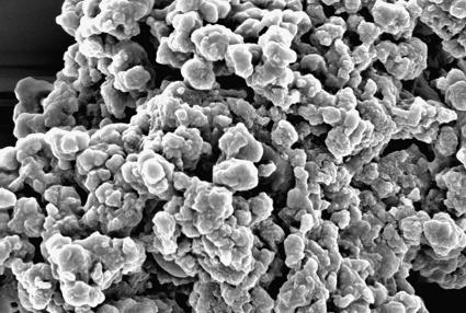
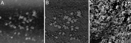
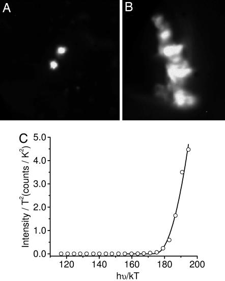
Neuromelanin (NM) isolated from the substantia nigra region of the human brain was studied by scanning probe and photoelectron emission microscopies. Atomic force microscopy reveals that NM granules are comprised of spherical structures with a diameter of approximately 30 nm, similar to that observed for Sepia cuttlefish, bovine eye, and human eye and hair melanosomes. Photoelectron microscopy images were collected at specific wavelengths of UV light between 248 and 413 nm, using the spontaneous-emission output from the Duke OK-4 free electron laser. Analysis of the data establishes a threshold photoionization potential for NM of 4.5 +/- 0.2 eV, which corresponds to an oxidation potential of -0.1 +/- 0.2 V vs. the normal hydrogen electrode (NHE). The oxidation potential of NM is within experimental error of the oxidation potential measured for human eumelanosomes (-0.2 +/- 0.2 V vs. NHE), despite the presence of a significant fraction of the red pigment, pheomelanin, which is characterized by a higher oxidation potential (+0.5 +/- 0.2 V vs. NHE). Published kinetic studies on the early chemical steps of melanogenesis show that in the case of pigments containing a mixture of pheomelanin and eumelanin, of which NM is an example, pheomelanin formation occurs first with eumelanin formation predominantly occurring only after cysteine levels are depleted. Such a kinetic model would predict a structural motif with pheomelanin at the core and eumelanin at the surface, which is consistent with the measured surface oxidation potential of the approximately 30-nm constituents of NM granules.
Conflict of interest statement
The authors declare no conflict of interest.
Figures
Comment in
-
Encapsulation of a reactive core in neuromelanin.Proc Natl Acad Sci U S A. 2006 Oct 3;103(40):14647-8. doi: 10.1073/pnas.0606879103. Epub 2006 Sep 27. Proc Natl Acad Sci U S A. 2006. PMID: 17005730 Free PMC article. No abstract available.
References
-
- Graham DG. Arch Pathol Lab Med. 1979;103:359–362. - PubMed
-
- Zucca FA, Giaveri G, Gallorini M, Albertini A, Toscani M, Pezzoli G, Lucius R, Wilms H, Sulzer D, Ito S, et al. Pigment Cell Res. 2004;17:610–617. - PubMed
-
- Zecca L, Fariello R, Riederer P, Sulzer D, Gatti A, Tampellini D. FEBS Lett. 2002;510:216–220. - PubMed
-
- Fenichel GM, Bazelon M. Neurology. 1968;18:817–820. - PubMed
Publication types
MeSH terms
Substances
LinkOut - more resources
Full Text Sources
Other Literature Sources
