. 2006 Oct;128(4):473-85.
doi: 10.1085/jgp.200609619.

Cyclic nucleotide-gated ion channels in rod photoreceptors are protected from retinoid inhibition

Affiliations

Cyclic nucleotide-gated ion channels in rod photoreceptors are protected from retinoid inhibition

Quanhua He et al. J Gen Physiol. 2006 Oct.

Abstract

In vertebrate rods, photoisomerization of the 11-cis retinal chromophore of rhodopsin to the all-trans conformation initiates a biochemical cascade that closes cGMP-gated channels and hyperpolarizes the cell. All-trans retinal is reduced to retinol and then removed to the pigment epithelium. The pigment epithelium supplies fresh 11-cis retinal to regenerate rhodopsin. The recent discovery that tens of nanomolar retinal inhibits cloned cGMP-gated channels at low [cGMP] raised the question of whether retinoid traffic across the plasma membrane of the rod might participate in the signaling of light. Native channels in excised patches from rods were very sensitive to retinoid inhibition. Perfusion of intact rods with exogenous 9- or 11-cis retinal closed cGMP-gated channels but required higher than expected concentrations. Channels reopened after perfusing the rod with cellular retinoid binding protein II. PDE activity, flash response kinetics, and relative sensitivity were unchanged, ruling out pharmacological activation of the phototransduction cascade. Bleaching of rhodopsin to create all-trans retinal and retinol inside the rod did not produce any measurable channel inhibition. Exposure of a bleached rod to 9- or 11-cis retinal did not elicit channel inhibition during the period of rhodopsin regeneration. Microspectrophotometric measurements showed that exogenous 9- or 11-cis retinal rapidly cross the plasma membrane of bleached rods and regenerate their rhodopsin. Although dark-adapted rods could also take up large quantities of 9-cis retinal, which they converted to retinol, the time course was slow. Apparently cGMP-gated channels in intact rods are protected from the inhibitory effects of retinoids that cross the plasma membrane by a large-capacity buffer. Opsin, with its chromophore binding pocket occupied (rhodopsin) or vacant, may be an important component. Exceptionally high retinoid levels, e.g., associated with some retinal degenerations, could overcome the buffer, however, and impair sensitivity or delay the recovery after exposure to bright light.


Figures

Figure 1.
Figure 1.
Inhibition of native rod channels by ATR. (A) Decrement in current in a membrane patch excised from a salamander rod outer segment after exposure to ATR. The minimum exposure was ∼1 h at each concentration to allow inhibition to reach steady state. Membrane voltage was stepped from a holding potential of 0 to ±100 mV in 50-mV increments at a saturating cGMP concentration. Currents measured in the absence of cGMP were subtracted from each trace. (B) Time course of inhibition. In another membrane patch, the fractional current in saturating cGMP declined exponentially with a time constant of 15 min due to the presence of 1.0 μM ATR. (C) The dose–response relation for ATR. Symbols plot the fractional inhibition of the steady-state, cGMP-activated currents at 100 mV from nine patches. A fit of the results with 1 − r/rmax = [ATR]n/(IC50 n + [ATR]n) yielded an IC50 of 250 nM and a Hill coefficient of 1.5.
Figure 2.
Figure 2.
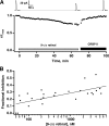
9-cis retinal–induced closure of channels in intact rods. (A) Slow decline in circulating current of a rod induced by 3 μM 9-cis retinal. Most of the circulating current was restored relatively rapidly after removal of the retinoids by extracellular perfusion with 0.3 μM CRBP II. Representative responses to saturating flashes are shown above (left to right): before treatment, after perfusion with 9-cis retinal for 1 h, and after washing with CRBP II. (B) Dependence of channel inhibition on [9-cis retinal]. Inhibition was assessed in 27 rods after a 60-min exposure to 9-cis retinal where each rod was only exposed to a single concentration of retinal. Linear regression of fractional inhibition against the logarithm of [9-cis retinal] (continuous line, r = 0.71) suggested a 0.12 loss in current per concentration decade.
Figure 3.
Figure 3.
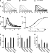
Averaged responses of a rod to flashes A, in darkness; B, in dim steady light; and C, in darkness after perfusion with 9-cis retinal to achieve the same reduction in circulating current as produced by background light. Responses to trials before and after exposure to a background light were indistinguishable so they were combined. The intensity of the background light was 0.024 photons μm−2 at 500 nm. Flashes were given at time = 0 s. (D) Little effect of 9-cis retinal on dim flash response kinetics. Flashes eliciting responses that were <20% of the maximal response were considered to be dim. Response kinetics were accelerated markedly by background light (gray trace) compared with the baseline condition (thin black trace), but not by retinoid treatment (bold, black trace). Responses were normalized by their peak amplitudes. (E) Lack of sensitivity change after 9-cis retinal treatment (baseline, open diamonds; 9-cis retinal treatment, black circles) in the rod whose responses are shown in A–C. Values for i1/2, the flash strength giving rise to a half-maximal response, were 4 and 5 photons μm−2 for baseline and retinal-treated conditions, respectively. Dim background light did reduce sensitivity several-fold, shifting the stimulus–response relation (gray triangles) to the right. Continuous lines show fits with an empirical function: r/rmax = 1 − exp(−k1 + k2exp)−k3i))i) where k1, k2, and k3 are constants, as described in Ma et al. (2001). Averaged values from seven rods for F, dim flash response time to peak; G, dim flash response integration time; and H, i1/2. Relative sensitivity is inversely proportional to i1/2. Error bars show the SEM.
Figure 4.
Figure 4.
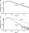
Lack of an increase in PDE activity after 11-cis retinal–induced loss of circulating current. Normalized current during steps into Li+ solution is plotted on semilogarithmic coordinates. The relative PDE rates are given by the ratio of the slopes of the falling phases of current decline. Time zero in the plot is the time at which the movement of the cell toward the Li+ solution interface was initiated. The delay between this time and the start of the current change is accounted for by the time necessary for the outer segment to reach this solution interface. (A) Measurements of PDE rates made before (thin line) and after (thick line) a 60-min exposure to 25 μM 11-cis retinal that reduced circulating current by a factor of 0.18. (B) Increased PDE rate in a different rod after exposure to bright light that bleached a fraction of the rhodopsin and reduced the circulating current by a factor of 0.19 (thick gray line), compared with the prebleached condition (thin black line).
Figure 5.
Figure 5.
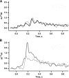
Estimation of guanylate cyclase activity. The maximum of the first derivative of the cube root of the normalized current, d(J1/3)/dt from a rod during steps into 0.5 mM IBMX solution provides an estimate of the relative GC rates. The solution change was initiated as described in the legend of Fig. 4. (A) Similar rates of cGMP synthesis before (thin line) and after (thick line) 60 min of treatment with 25 μM 11-cis retinal that resulted in 0.15 inhibition of the circulating current. (B) 2.5-fold increase in the rate of cGMP synthesis in another rod after a fractional bleach of its rhodopsin that reduced the circulating current at steady state by a factor of 0.23 (prebleach, thin black line; postbleach, thick gray line).
Figure 6.
Figure 6.
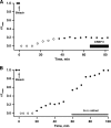
Absence of channel inhibition by retinoid after pigment bleaching. (A) The ordinate plots the amplitude of the response to a fixed flash strength, relative to the amplitude at the beginning of the recording. Saturating responses are plotted with filled symbols, while subsaturating responses or trials in which no response was observed were plotted with open symbols. Exposure to 3.5 × 10⁸ photons μm−2 at 500 nm at time = 0 min to bleach 91% of the rhodopsin eliminated the circulating current and desensitized the rod. Circulating current and sensitivity partially recovered after ∼40 min. No additional circulating current was obtained upon perfusion with 0.3 μM CRBP II to remove retinoids. (B) A second rod was exposed to 2.7 × 10⁸ photons μm−2 at 500 nm at time = 0 min to bleach 84% of the rhodopsin. 59.3 min later, the rod was perfused with 4 μM 9-cis retinal to regenerate the rhodopsin. The circulating current recovered completely to the prebleached level.
Figure 7.
Figure 7.
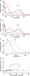
Microspectrophotometric measurements of retinoid uptake into outer segments that were attached to (n = 2 rods), A, or had detached from (n = 3 ROSs), B, their inner segments.Measurements were made before (black lines) and after (red lines) treatment with 9-cis retinal for ∼4 h with the probe beam polarized parallel (dashed lines) and perpendicular (continuous lines) to the outer segment axis. The ROSs in B were also treated with CRBPII for 2.5 h. Nevertheless, their absorbance at 370 nm was the highest of any ROS measured. Spectra from untreated outer segments were scaled by their absorbance at 280 nm. Spectra from treated outer segments were scaled so that the absorbance at 520 nm would match that of the corresponding untreated spectra. Absorbance increased at 325 nm in outer segments retaining an inner segment and at 370 nm in outer segments lacking an inner segment. (C) Normalized spectra from rods and ROSs, taken as the difference between the measurement with and without exposure to 9-cis retinal for the probe beam polarized parallel to the rod axis. (D) Slow, variable uptake of 9-cis retinal by rods. Measurements on 25 intact rods were made with the probe beam polarized perpendicular to the long axis of the outer segment. Normalized retinol content was determined by decomposition of the spectra of individual rods into A1 and A2 pigment components (Govardovskii et al., 2000), retinal, retinol, and the protein band absorbing at 502, 521, 373, 325, and 280 nm, respectively. Retinoid and protein absorption bands were assumed to be Gaussian in form. The amplitude of the retinol band was divided by that of the protein band to adjust for outer segment diameter. Serial results from the same rod were connected with a continuous line. Dashed lines demarcate the mean ± one standard deviation for 34 untreated rods.
Figure 8.
Figure 8.
Rapid regeneration of rhodopsin in a bleached rod with 11-cis retinal. Absorption spectra are shown before bleaching (continuous black line), after bleaching with bright light at 500 nm (dotted black line), and after the addition of 30 μM 11-cis retinal (thin red line, 22 min treatment; thick red line, 42 min treatment).

Similar articles

Cited by

References

    1. Allikmets, R., N. Singh, H. Sun, N.F. Shroyer, A. Hutchinson, A. Chidambaram, B. Gerrard, L. Baird, D. Stauffer, A. Peiffer, et al. 1997. A photoreceptor cell-specific ATP-binding transporter gene (ABCR) is mutated in recessive Stargardt macular dystrophy. Nat. Genet. 15:236–246. - PubMed
    1. Brown, P.K., and G. Wald. 1956. The neo-b isomer of vitamin A and retinene. J. Biol. Chem. 222:865–877. - PubMed
    1. Brown, P.K., I.R. Gibbons, and G. Wald. 1963. The visual cells and visual pigment of the mudpuppy, Necturus. J. Cell Biol. 19:79–106. - PMC - PubMed
    1. Cohen, G.B., D.D. Oprian, and P.R. Robinson. 1992. Mechanism of activation and inactivation of opsin: role of Glu113 and Lys296. Biochemistry. 31:12592–12601. - PubMed
    1. Cornwall, M.C., G.J. Jones, V.J. Kefalov, G.L. Fain, and H.R. Matthews. 2000. Electrophysiological methods for measurement of activation of phototransduction by bleached visual pigment in salamander photoreceptors. Methods Enzymol. 316:224–252. - PubMed

Publication types

MeSH terms