. 2006 Dec;149(7):958-65.
doi: 10.1038/sj.bjp.0706913. Epub 2006 Sep 25.

Na+-K+-ATPase is involved in the sustained ACh-induced hyperpolarization of endothelial cells from rat aorta

Affiliations

Na+-K+-ATPase is involved in the sustained ACh-induced hyperpolarization of endothelial cells from rat aorta

A Bondarenko et al. Br J Pharmacol. 2006 Dec.

Abstract

Background and purpose: Inhibition of Na(+)-K(+)-ATPase is known to attenuate endothelium-dependent relaxation in many arteries. The purpose of this study was to evaluate the role of Na(+)-K(+)-ATPase in the regulation of endothelial membrane potential at rest and during stimulation by ACh.

Experimental approach: Membrane potential was recorded from the endothelium of rat aorta using the perforated patch-clamp technique.

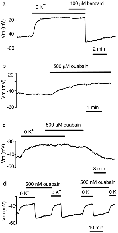
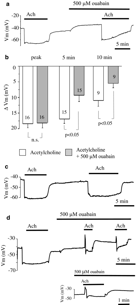
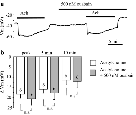
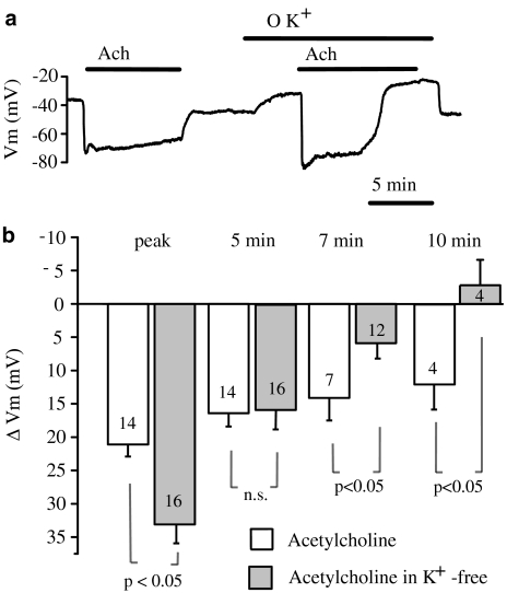
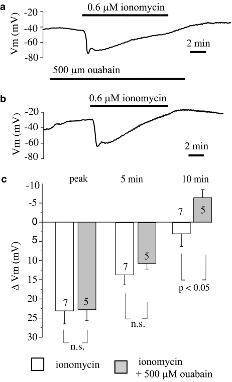
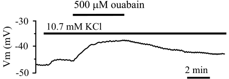
Key results: Superfusion with K(+)-free solution produced a depolarization of about 11 mV from the resting value of -42.9+/-0.9 mV. Reintroduction of 4.7 mM K(+) transiently hyperpolarized endothelial cells to -52.4+/-1.8 mV and the membrane potential recovered within 10 min. Ouabain 500 microM depolarized endothelium by about 11 mV and inhibited the hyperpolarization induced by K(+) reintroduction into the K(+)-free solution. However, 500 nM ouabain did not affect the resting membrane potential or the hyperpolarization induced by K(+) reintroduction. Pre-exposure to ouabain 500 microM, but not 500 nM, attenuated the sustained component of hyperpolarization to ACh without affecting the amplitude of the transient peak hyperpolarization. In K(+)-free solution, the amplitude of peak hyperpolarization to ACh was increased, while the sustained component of hyperpolarization was attenuated.

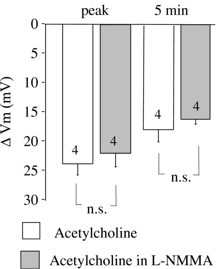
Conclusions and implications: These results indicate that electrogenic Na(+)-K(+)-ATPase partially contributes to the sustained hyperpolarization of endothelial cells from rat aorta in response to ACh. They also suggest that the alpha1, but not alpha2 or alpha3 isoforms, is involved in ACh-mediated hyperpolarization.


Figures

Figure 1
Figure 1
Modulation of endothelial membrane potential by Na+-K+-ATPase inhibition. (a) Effects of external K+ withdrawal followed by 100 μM benzamil administration and K+ reintroduction on the membrane potential. (b) Effect of 500 μM ouabain on endothelial membrane potential. (c) Lack of depolarizing effect of 500 μM ouabain in the absence of external K+ and inhibitory effect of 500 μM ouabain on the hyperpolarization induced by K+ reintroduction. (d) Lack of inhibitory effect of 500 nM ouabain on the hyperpolarization induced by K+ reintroduction.
Figure 2
Figure 2
Effect of 500 μM ouabain on the hyperpolarization of endothelial cells from rat aorta produced by acetylcholine. (a) Original tracing showing the influence of 500 μM ouabain on the hyperpolarization of endothelial cells produced by 2 μM acetylcholine. (b) Graphical representation of the time dependency of the effect of 500 μM ouabain on the mean changes in membrane potential evoked by 2 μM acetylcholine. The changes in membrane potential were calculated as the difference between the membrane potential values before and after acetylcholine administration. Numbers associated with bar plot indicate the number of aorta preparations. (c) Representative record of repeated applications of 2 μM acetylcholine. (d) Original tracing showing attenuation of all phases of acetylcholine-evoked hyperpolarization by 500 μM ouabain that unmasked a transient depolarization phase in response to acetylcholine. For clarity, a portion of the trace has been expanded.
Figure 3
Figure 3
Influence of a low concentration of ouabain (500 nM) on the hyperpolarization of endothelial cells produced by acetylcholine. (a) Original tracing showing a lack of effect of 500 nM ouabain on the hyperpolarization of endothelial cells produced by 2 μM acetylcholine. (b) Graphical representation of the time dependency of the effect of 500 nM ouabain on the mean changes in membrane potential evoked by 2 μM acetylcholine. The changes in membrane potential were calculated as the difference between the membrane potential values before and after acetylcholine administration. Numbers associated with bar plot indicate the number of aorta preparations.
Figure 4
Figure 4
Effect of K+-free solution on the hyperpolarization of endothelial cells from rat aorta produced by acetylcholine. (a) Original tracing showing the influence of external K+ substitution on the resting membrane potential and acetylcholine-induced hyperpolarization of endothelial cells. (b) Graphical representation of the time dependency of the effect of K+-free solution on the mean changes in membrane potential evoked by 2 μM acetylcholine. The changes in membrane potential were calculated as the difference between the membrane potential values before and after acetylcholine administration. Numbers associated with bar plot indicate the number of aorta preparations.
Figure 5
Figure 5
Influence of 500 μM ouabain on the hyperpolarization of endothelial cells from rat aorta produced by ionomycin. (a) Original tracing of an experiment showing the influence of ionomycin (0.6 μM) on the membrane potential of endothelial cells. (b) Original tracing of an experiment showing the influence of 500 μM ouabain on the hyperpolarization of endothelial cells produced 0.6 μM ionomycin. (c) Graphical representation of the time dependency of the effect of 500 μM ouabain on the mean changes in membrane potential evoked by 0.6 μM ionomycin. The changes in membrane potential were calculated as the difference between the membrane potential values before and after ionomycin administration. Numbers associated with bar plot indicate the number of aorta preparations.
Figure 6
Figure 6
Original tracing of the membrane potential responses to increased KCl (from 4.7 to 10.7 mM) followed by 500 μM ouabain administration.
Figure 7
Figure 7
Effect of NO synthase inhibitor L-NMMA (200 μM) on the mean changes in membrane potential of endothelial cells evoked by 2 μM acetylcholine. The changes in membrane potential were calculated as the difference between the membrane potential values before and after acetylcholine administration. Numbers associated with bar plot indicate the number of aorta preparations.

References

    1. Blanco G, Mercer RW. Isozymes of the Na,K-ATPase: heterogeneity in structure, diversity in function. Am J Physiol. 1998;275:F633–F650. - PubMed
    1. Blaustein MP, Lederer WJ. Sodium/calcium exchange: its physiological implications. Physiol Rev. 1999;79:763–854. - PubMed
    1. Bondarenko A. Sodium–calcium exchanger contributes to membrane hyperpolarization of intact endothelial cells from rat aorta during acetylcholine stimulation. Br J Pharmacol. 2004;143:9–18. - PMC - PubMed
    1. Bray K, Quast U. Differences in the K+-channels opened by cromakalim, acetylcholine and substance P in rat aorta and porcine coronary artery. Br J Pharmacol. 1991;102:585–594. - PMC - PubMed
    1. Brayden JE. Membrane hyperpolarization is a mechanism of endothelium-dependent cerebral vasodilation. Am J Physiol. 1990;259:H668–H673. - PubMed

MeSH terms

LinkOut - more resources