. 2006 Nov;149(5):490-7.
doi: 10.1038/sj.bjp.0706909. Epub 2006 Sep 25.

RANTES stimulates Ca2+ mobilization and inositol trisphosphate (IP3) formation in cells transfected with G protein-coupled receptor 75

Affiliations

RANTES stimulates Ca2+ mobilization and inositol trisphosphate (IP3) formation in cells transfected with G protein-coupled receptor 75

A Ignatov et al. Br J Pharmacol. 2006 Nov.

Abstract

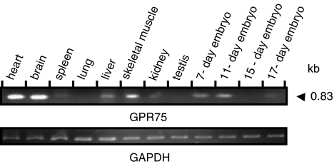
Background and purpose: RANTES is an inflammatory chemokine with a critical role in T-lymphocyte activation and proliferation. Its effects are mediated through G protein-coupled heptahelical receptors (GPCRs). We show for the first time that RANTES activates the orphan G protein-coupled receptor 75 (GPR75).

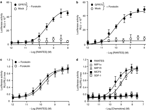
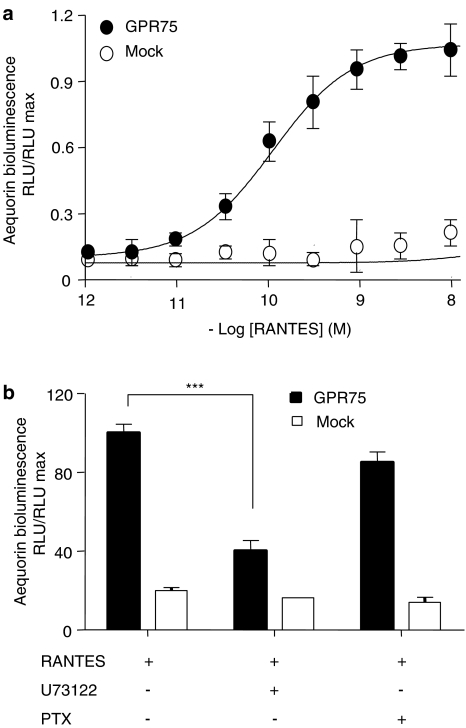
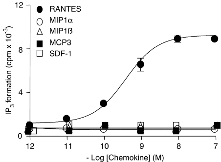
Experimental approach: To identify a ligand for GPR75 we have used three different and independent methods, namely luciferase assay, bioluminescence assay and IP3 accumulation assay.

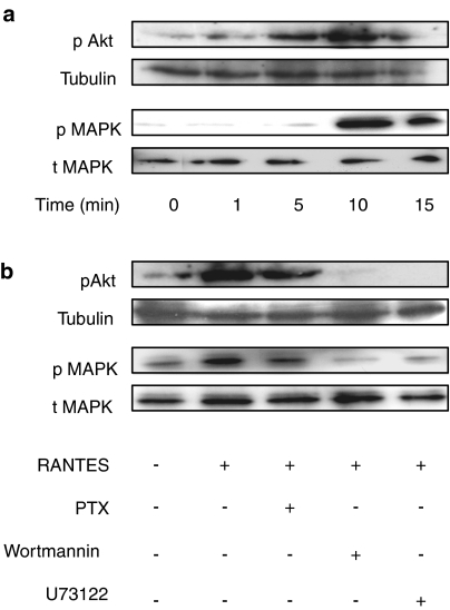
Key results: Treatment of cells expressing GPR75 with subnanomolar concentrations of RANTES led to stimulation of the luciferase activity in a reporter-gene assay, an increase in inositol trisphosphate, and intracellular Ca2+. The latter effect was blocked by the phospholipase-C inhibitor (PLC) U73122 indicating that Gq proteins mediate GPR75 signaling. RANTES enhanced the phosphorylation of AKT and mitogen-activated protein kinase (MAPK) in GPR75-transfected cells and this effect was blocked by the PLC inhibitor U73122 and the phosphatidylinositol 3-kinase (PI3K) inhibitor, wortmannin. The hippocampal cell line HT22, which expresses GPR75 endogenously, but not the other known RANTES receptors, was used to study the effects of RANTES and GPR75 on neuronal survival. Treatment of HT22 cells with RANTES significantly reduced the neurotoxicity of amyloid-beta peptides, by activating PLC and PI3K.

Conclusions and implications: This demonstrate clearly and undoubtedly the ability of RANTES to act on GPR75. Defects in the RANTES/GPR75-signaling pathway may contribute to neuroinflammatory and neurodegenerative processes as observed in Alzheimer's disease.


Figures

Figure 1
Figure 1
Expression analysis of mouse GPR75 mRNA. The expression of GPR75 mRNA in different adult mouse tissues and 7-, 11-, 15- and 17-day-old embryos was assessed by PCR using a multiple-tissue cDNA panel and GPR75-specific primers that amplify a product of 843 bp (upper panel). GAPDH served as loading control (bottom panel).
Figure 2
Figure 2
GPR75-mediated stimulation of luciferase expression by RANTES. CV-1 cells were transiently transfected with either GPR75 or empty vector (Mock) and the MRE/SRE/CRE-reporter construct. 48 h later, cells were stimulated for 5 h with different concentrations of RANTES in the absence (a), or presence (b) of 5 μM forskolin. The luciferase activity is expressed in relative light units (RLU). (c) The stimulation of the luciferase activity given as a ratio between the luminescence measured for the individual samples in RLU and the maximal stimulation (RLU max). The calculated EC50 values were: 0.11 and 0.16 nM. (d) Cells were stimulated for 5 h with 5 μM forskolin and with various concentrations of different chemokines. Note that in addition to RANTES, only MIP1α increased the luciferase activity over the forskolin-stimulated background response with an EC50 value of 1 nM. Data are expressed as mean values±s.d.
Figure 3
Figure 3
RANTES stimulates the Ca2+-induced bioluminescence in GPR75-transfected CHO-K1 cells. (a) CHO-K1 cells were transiently co-transfected with GPR75 or empty vector and apoaequorin as Ca2+ sensor. Cells were treated with different concentrations of RANTES, and the Ca2+-induced bioluminescence was measured at 469 nm and is expressed as RLU/RLU max. The dose–response curve yielded an EC50 value of 0.12 nM. (b) CHO-K1 cells transiently co-transfected with apoaequorin and GPR75 or empty vector were pretreated with 50 ng ml−1 PTX for 18 h, with 5 μM PLC inhibitor U73122 for 10 min, before the response to RANTES was measured as Ca2+-induced luminescence. The RANTES-induced Ca2+ increase was significantly inhibited by U73122 compared to untreated controls, whereas PTX had no effect (***P<0.001). Data are expressed as mean values±s.d.
Figure 4
Figure 4
RANTES stimulates IP3 formation in GPR75-transfected HEK293 cells. HEK293 cells were transiently transfected with GPR75. Cells were treated with various concentrations of RANTES, MIP1α, MIP1β, MCP3 and SDF-1 for 30 min, and the production of IP3 was measured in extracts from intact HEK293 cells. The dose–response curve yielded an EC50 value of 0.3 nM for RANTES-induced production of IP3. Data are expressed as mean values±s.d.
Figure 5
Figure 5
RANTES induces the phosphorylation of Akt and MAPK in GPR75-transfected CV-1 cells via the PLC/PI3K/Akt signaling pathway. (a) CV-1 cells were transiently transfected with GPR75 and 48 h later incubated with 1 nM RANTES for the indicated times. Whole-cell extracts were analyzed by Western blotting using antibodies against phosphorylated MAPK (pMAPK), phosphorylated Akt (pAkt), total MAPK (tMAPK) and acetylated tubulin. Note that the levels of total MAPK and tubulin remained constant. The blot is a representative of three different experiments. (b) GPR75-transfected CV-1 cells were pretreated with 50 ng ml−1 PTX for 18 h, with 5 μM PLC inhibitor U73122 for 10 min, and with 0.5 μM PI3K inhibitor wortmannin for 1 h, before1 nM RANTES was added for 10 min. The blot is representative of two experiments.
Figure 6
Figure 6
Aβ peptides reduce cell viability in HT22 cells. (a) HT22 cells express GPR75 endogenously but not CCR1, CCR3 and CCR5 chemokine receptors (upper panel). GAPDH served as a loading control (lower panel). cDNA represents the negative control. The experiment is representative of three detections. (b) HT22 cells respond to a 24 h treatment with Aβ peptides by cell death, as assessed by the MTT assay. Note that no difference was observed between 20 μM Aβ1−42 and Aβ25−35, whereas Aβ42−1 had no effect. Data are expressed as mean values±s.d.
Figure 7
Figure 7
RANTES protects HT22 cells from cell death induced by Aβ peptide. (a) HT22 cells were preincubated for 1 h with and without 1 nM RANTES (R), before addition of 20 μM Aβ1−42 (Aβ). After 24 h, cell viability was determined using the MTT assay. The effect of RANTES on cell survival was significant compared to cells treated with Aβ alone (*P<0.05). Additional transient transfection with GPR75 increased the protective effect of RANTES (**P<0.01). The protective effect of RANTES was significantly inhibited by pretreatment of HT22 cells for 1 h with 0.5 μM wortmannin (W) or for 10 min with 5 μM U73122 (U), before RANTES was added (*P<0.05). (b and c) HT22 cells were seeded in six-well plates for 24 h. They were incubated for 24 h with 20 μM Aβ1−42 without (b) or with (c) preincubation with 1 nM RANTES for 1 h and visualized by phase-contrast microscopy. Scale bar – 50 μm. (d) After pretreatment for 1 h with 1 nM of the indicated chemokines, HT22 cells were incubated with 20 μM Aβ25−35, and after 24 h the cell viability was determined by the MTT assay. Only RANTES and, to a lesser extent, MIP1α had a significant neuroprotective effect (**P<0.01 and *P<0.05, respectively). The data in (a) and (d) are expressed as mean values±s.d.

Comment in

References

    1. Bajetto A, Bonavia R, Barbero S, Florio T, Schettini G. Chemokines and their receptors in the central nervous system. Front Neuroendocrinl. 2001;22:147–184. - PubMed
    1. Cartier L, Hartley O, Dubois-Dauphin M, Krause KH. Chemokine receptors in the central nervous system: role in brain inflammation and neurodegenerative diseases. Brain Res Brain Res Rev. 2005;48:16–42. - PubMed
    1. Crane IJ, Kuppner MC, McKillop-Smith S, Knott RM, Forrester JV. Cytokine regulation of RANTES production by human retinal pigment epithelial cells. Cell Immunol. 1998;184:37–44. - PubMed
    1. Cummings JL, Vinters HV, Cole GM, Khachaturian ZS.Alzheimer's disease: etiologies, pathophysiology, cognitive reserve, and treatment opportunities Neurology 199851S2–S17.(Discussion S65–S17) - PubMed
    1. Hirata A, Masuda S, Tamura T, Kai K, Ojima K, Fukase A, et al. Expression profiling of cytokines and related genes in regenerating skeletal muscle after cardiotoxin injection: a role for osteopontin. Am J Pathol. 2003;163:203–215. - PMC - PubMed

MeSH terms